Interference with the EKG reading is known as artifact and it can come from a variety of both patient and non-patient factors. The most common reasons for artifact caused by the patient include:
seizures
fast breathing
wet skin
and shivering
Non-patient causes include use of electrodes with dry gel, damaged patient cables, electromagnetic frequency, and interference from cell phones and other medical and non-medical devices. Artifact can make impossible for the doctor to interpret the rhythm.
Wet skin:
Some patients will be diaphoretic during EKG acquisition. The technician
can wipe the patient off with a towel or clean the patient's skin with an alcohol pad before placing the electrodes. The alcohol needs to completely dry prior to applying the EKG electrodes.
Dry gel:
The gel on the EKG electrodes is specially designed to interface with the
patient's skin. The gel requires full surface area contact with the patients skin. Electrodes with dry gel should not be used.
Some newer EKG machine can detect artifact and correct for it but recognizing and eliminating artifacts is still and essential skill for the EKG technician.
Types of artifacts:
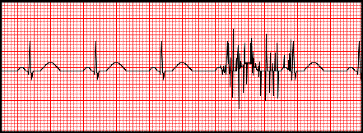
Somatic Tremors or muscle tremors:
Muscle movement on the EKG tracing is referred to somatic tremor. Somatic tremor exhibits as uneven spikes in the EKG tracings.
Causes of Somatic Tremors:
Seizure:
Seizure activity will cause huge artifact problems on the EKG due to uncontrolled muscle movement. Seizure activity must be controlled prior to acquiring the EKG tracing.
Trembling:
Some patients may be anxious or cold. The EKG technician must reassure the patient to keep him/her calm, provide warm blankets to control shivering, and attempt to move the electrodes to an area with minimal tremor.
Patients who have conditions such as Parkinson’s Disease are prone to uncontrolled muscular movement. Placing their palms in a prone position and under the buttocks will help reduce these interferences.
Patient movement:
Some patients might unconsciously be moving their legs or fidgeting with their hands during the test. Simply make the Patient aware of their movement and continue with procedure.
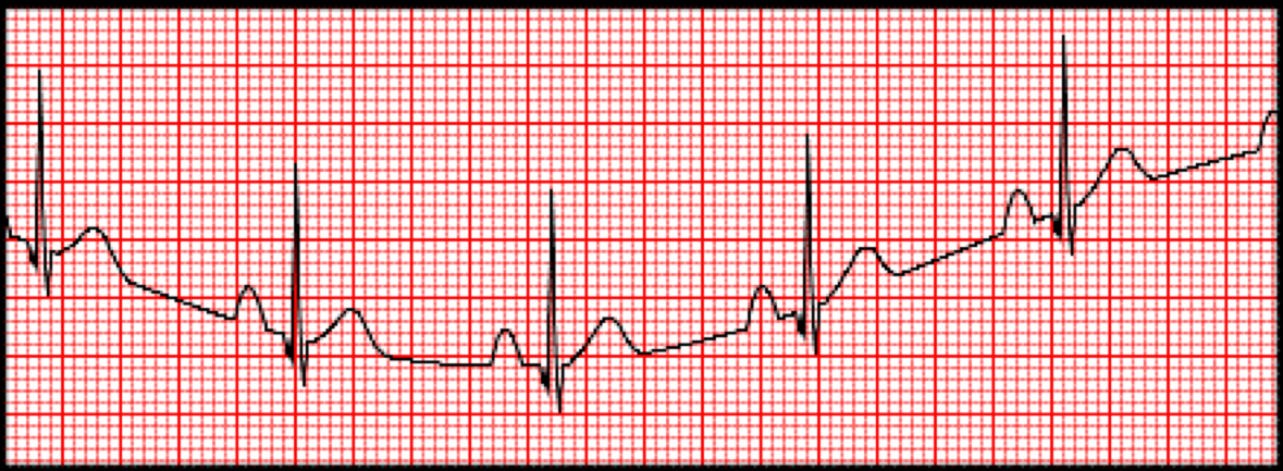
Wandering baseline:
A wandering baseline often appears on the EKG when the electrodes are improperly placed on the patient's torso.
Wandering Baseline Artifact can result from:
Movement of the cables or leads during the reading,
Loose electrodes
Dry electrodes
Labored breathing
Improper skin preparation
Leaving traces of lotion or oils on the skin
An easy way to reduce the wandering baseline artifact is to clean the skin where the electrodes are placed to remove lotion or oil, which causes a poor connection between the skin and the leads.
Another way to eliminate the wandering baseline is to move the limb leads to the wrists and ankles.
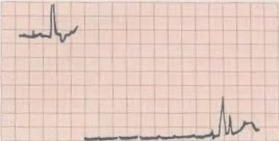
Interrupted baseline:
an interrupted baseline is demonstrated by a tracing that is not continuous.
A break in the baseline or a fully non-recorded lead can be seen.
Cause of interrupted baseline artifact:
Loose and dirty electrodes
Broken leads
Disconnected leads
Regular observation and maintenance of the lead wires, as well as double-checking connections prior to running the EKG, will help reduce this issue.
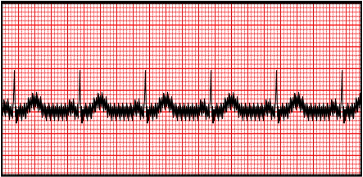
Electrical interference or 60 cycle interference:
Any electronic device on or near the patient can cause electrical interference.
This type of artifact is recognizing as uniform sharp spikes on the EKG tracing.
Causes of 60 cycle interference:
Cell phones
Smart watches
Any electrical device touching or near patient
EKG technician may need to use alternate electrodes placement in the following situations:
Medical or Cosmetic implants:
electrodes should not be placed over implants like a pacemaker, or over a breast implant. In this case the electrode should be placed as closely as possible to the stand position.
Large breast:
Move the breast tissue away from the electrode site as much as possible, to allow the placement of the electrodes.
Pregnancy:
If a patient is over six months pregnant, she cannot be placed in the supine position for an EKG.
Lying down in the supine position could cause the fetus to compress the patient’s vena cava and reduce blood return to the heart.
She will need to lie down and be tilted slightly( 15 degree ) to the left.
A patient with a history of respiratory or heart problems may be position in a semi fowler position. (laying with head elevated)