Chapter 7:
Rhythm Interpretation - Too Fast, Too Slow, Deadly
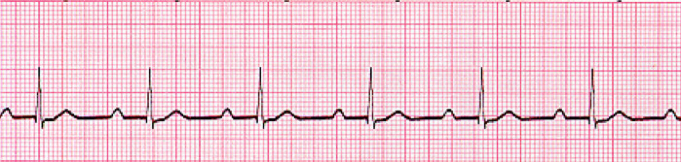
Normal Sinus Rhythm:
Normal sinus rhythm is 60-100x bpm. and shows all the following characteristics.
Use these five steps to analyze heart rhythms:
1) Is it Regular
2) heart rate between 60 and 100 bpm.
3) P to QRS Ratio
4) QRS interval of less than 0.12 seconds. (3 small boxes or less)
5) P-R interval between 0.12 and 0.20 seconds. (3 to 5 small boxes)
Too Fast, Too Slow:
Sinus Tachycardia: The heart rate is simply too fast.
All other characteristics are normal except for the heart rate greater than 100x minutes.
Most of the time, sinus tachycardia is a normal response of the cardiovascular system increasing the heart rate.
Tachycardia is normal during exercise or when people simply get tired.
Sinus bradycardia rhythm: The heart rate is slow, all other characteristics are normal except for a heart rate of less than 60 x min.
Sinus bradycardia is not necessarily abnormal. For some athletes, patient how have medical conditions, or older adults who live a basically sedentary lifestyle, this rate might be normal.
Signs and symptoms: syncope, dizziness, fatigue, shortness of breath, chest pain, confusion, or memory problems.
Treatment: pacemaker
Atrial Dysrhythmias:
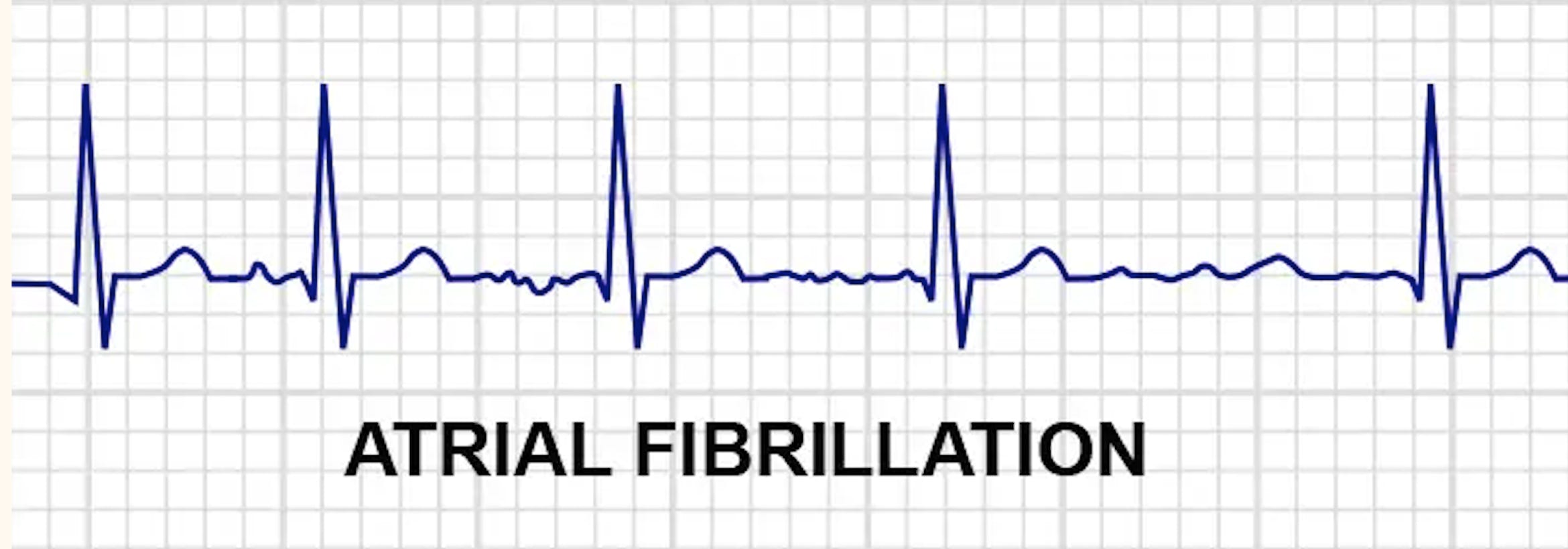
Atrial fibrillation: (often called A-fib) is a very common dysrhythmia , especially in older patients.
Atrial fibrillation makes the isoelectric line of the EKG tracing wavy and irregular with no clear P waves.
The QRS complex looks normal and has a normal duration, but the R-R interval is irregular.
Signs and symptoms: palpitations, weakness, fatigue, dizziness, shortness of breath chest pain.
Cause: hypertension, heart attack, coronary artery disease, abnormal heart valves.
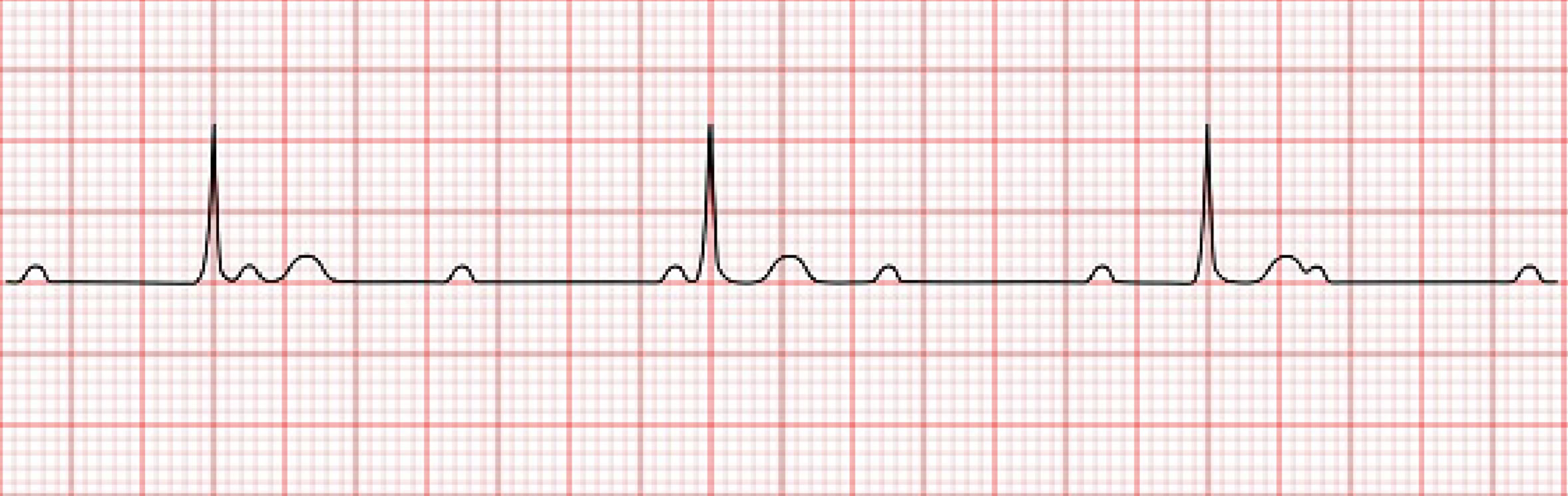
Atrial Flutter:
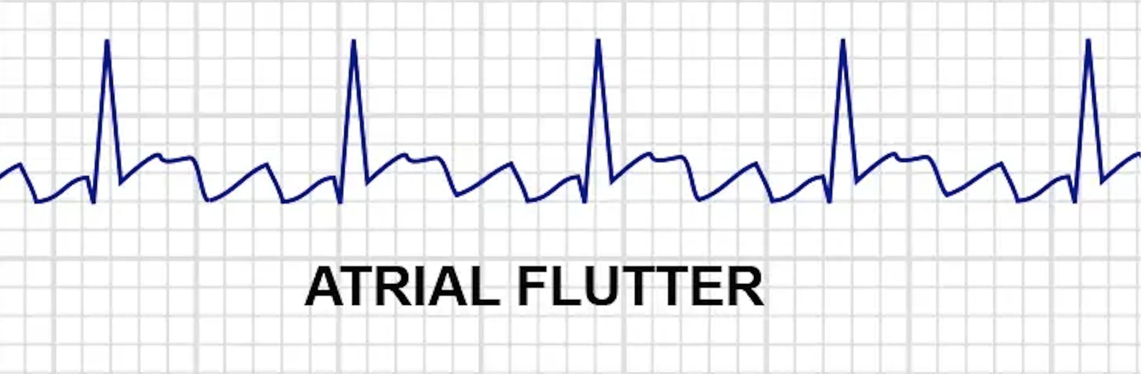
Atrial flutter: Is a condition in which the atria are contracting at a rate much faster than the ventricles are contracting.
The heart rate is fast, but usually with a regular rhythm.
The EKG tracing will show more P waves than QRS complex in the rhythm.
The P-R interval is not measurable in the atrial flutter, the QRS complex is usually normal.
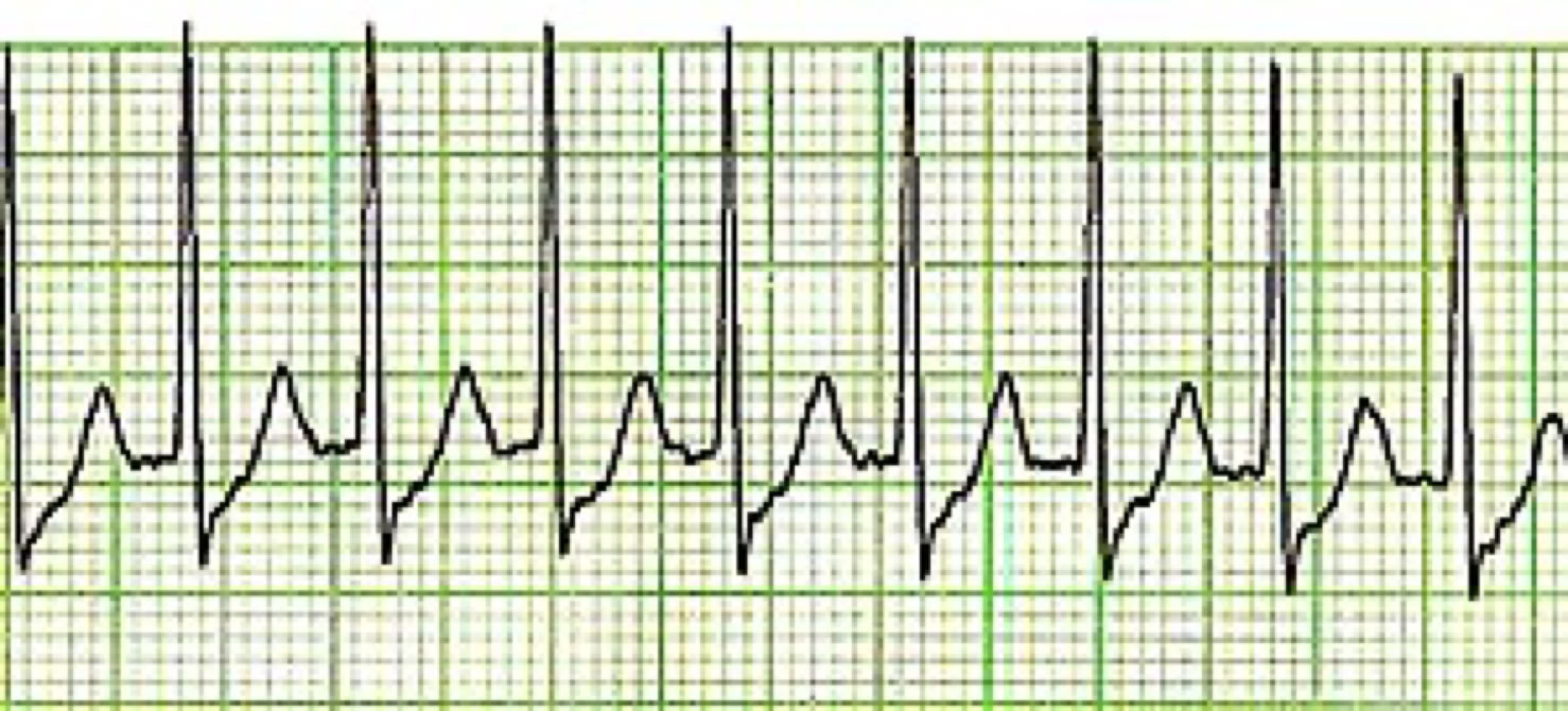
Supraventricular Tachycardia:
Supraventricular tachycardia or narrow complex tachycardia (SVT): In this situation, the impulse is not following the normal electrical conduction pathway.
Heart rate will be greater than 150/min, P waves are usually not visible.
Signs and symptoms: fluttering in your chest, palpitations, shortness of breath, dizziness, sweating, syncope.
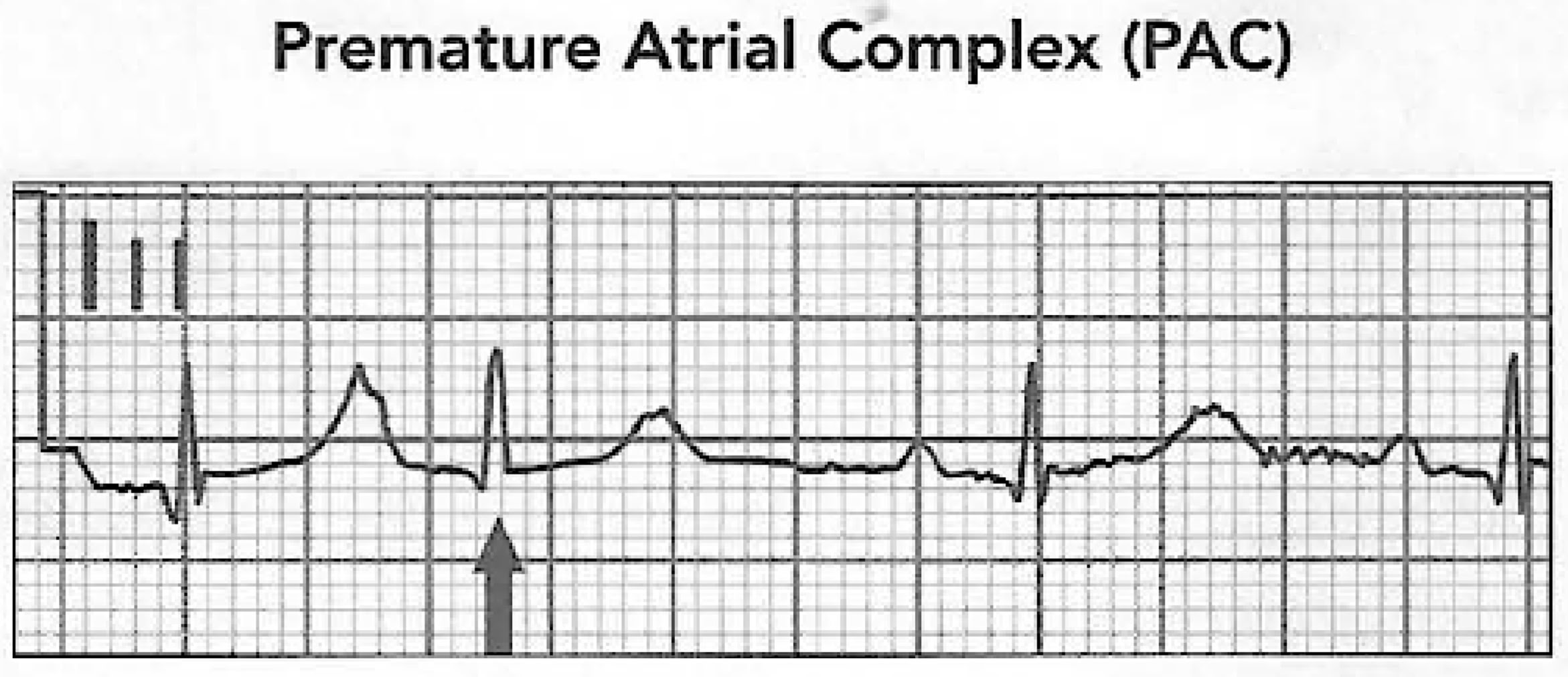
Premature Atrial Complex (PACs):
Premature atrial complex (PACS): are the result of electrical activity that originated in the atria.
The underlying rhythm is often normal but there are obvious early beats.
The QRS complex and the T wave in a premature atrial complex will be normal, but the P wave will not be normal, it may be buried in the preceding T wave, and the P-R interval may be shortened.
The P wave in these early beats may be taller and more pointed.
PACs are not rhythms! They are complexes that appear in an underlying rhythm.
First name the underlying rhythm, and then add PAC.
For example, the following tracing reveals regular sinus rhythm with one PAC.
The premature beat is the beat that occurs early in the cycle, followed by a rest period known as a compensatory pause.
This pause is a built-in delay mechanism enabling the heart to resume its electrical activity on time following a premature beat.
Heart Blocks:
Heart blocks are a special set of arrhythmias that indicate a difficulty in communication or no communication between the atria and ventricles.
In a normally functioning heart, the AV node and AV bundles slow down impulses from the SA node, allowing the ventricles time to fill with blood.
The severity depends on the location and cause of the blockage.
Heart blocks can usually be identified by a prolonged P-R interval.
First-degree heart block:
First-degree heart block represents a slow or delayed conduction through the AV node, resulting in a prolonged PR interval.
Specifically, the PR segment elongates in first-degree heart block.
P-R interval greater than 0.20sec
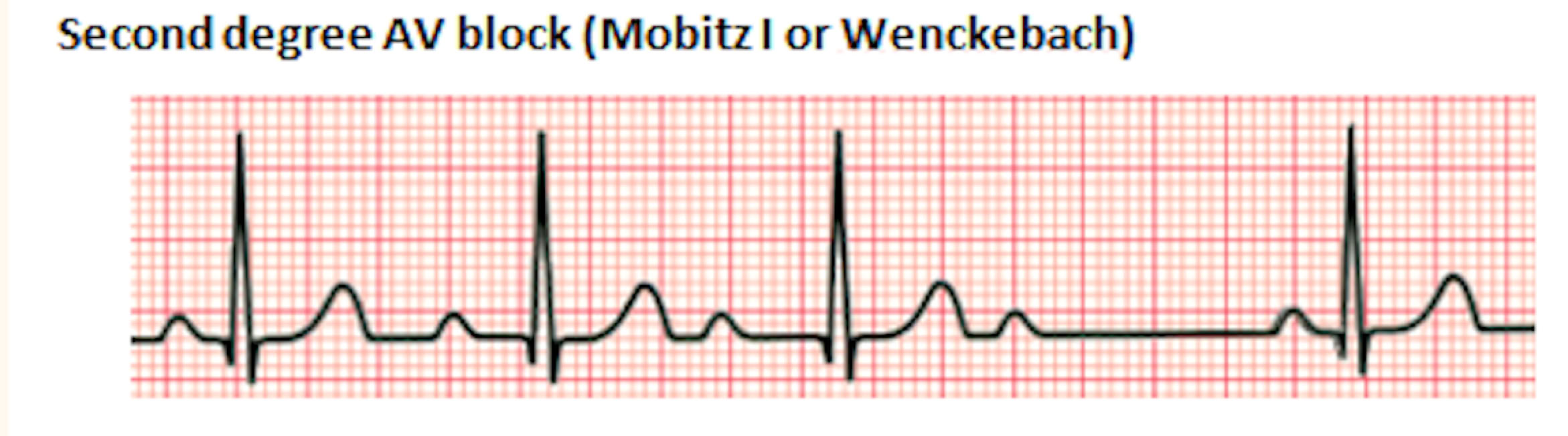
Second-degree Heart Block:
A second-degree heart block can occur in two forms:
Mobitz I (also sometimes called Wenckebach)
In this type of block, there are nonconductor or blocked impulses from the AV node to the ventricles.
As a result, there will be missing QRS complex. The P-R interval will get progressively longer until a QRS is missing, and then the patterns repeat itself.
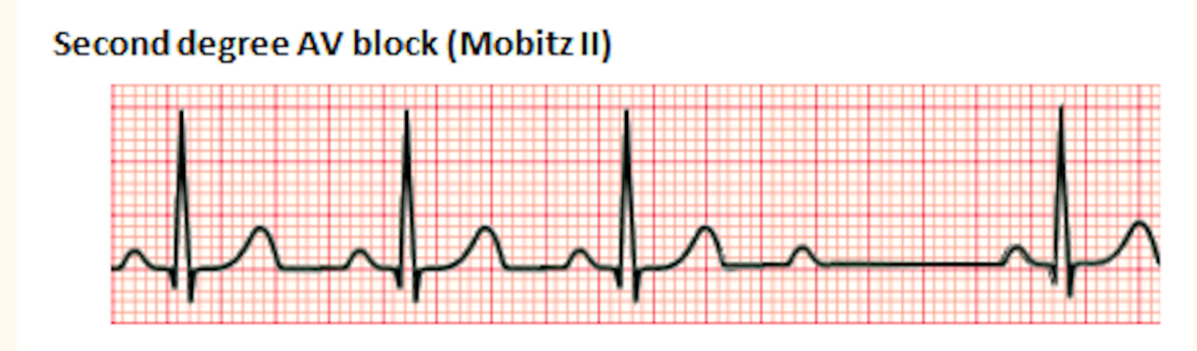
Mobitz type II (second-degree atrioventricular block, type II)
This is the classic form of heart block. The P-R interval remains constant, but the P wave is present with no QRS complex or T wave.
In this situation, the AV node has selectively blocked specific impulses. This type of heart block tends to progress quickly to complete heart block.
Both types of Mobitz have some impulses that are totally blocked by the AV node, but the pattern of the block differs.
In the Mobitz type I the P-R interval of the conducted beats get longer and longer until a beat is totally blocked.
while in Mobitz ii the P-R segment is normal.
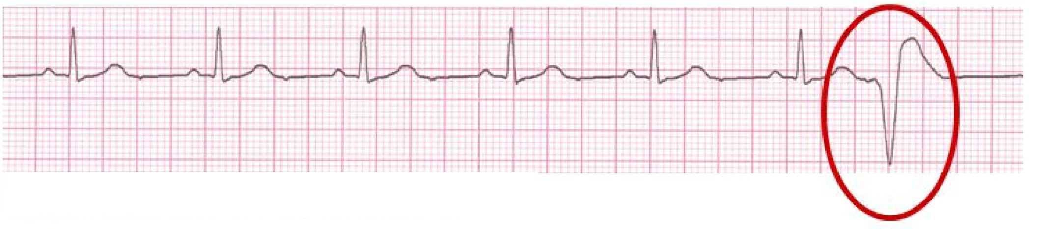
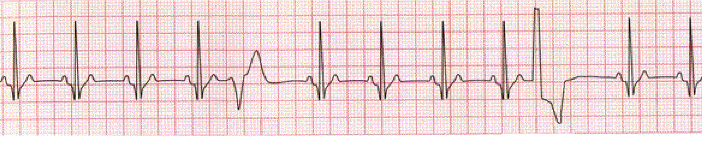
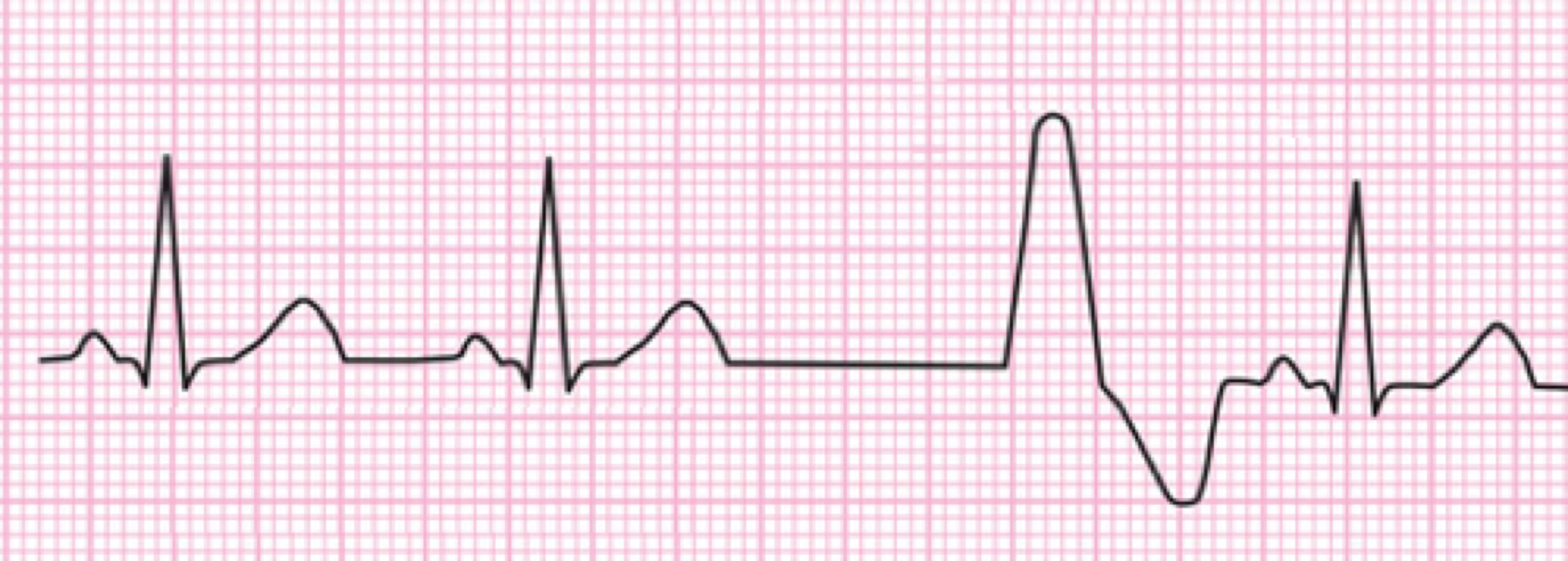
Premature Ventricular Complex (PVCs):
PVCs are ventricular depolarizations that occur early in the cardiac cycle. The characteristic findings on the EKG include:
• Absent P wave preceding the early QRS
• A wide QRS complex
• A QRS that looks different than the QRS complexes in the underlying rhythm
• The direction of the QRS complex and the T wave oppose one another
Like PACs, PVCs are not rhythms. They are complexes that appear in an underlying rhythm.
First name the underlying rhythm, then add PVC. For example, the tracing above reveals a regular sinus rhythm with PVCs.
PVCs can appear in many different patterns and with different shapes; in this case, the term "multifocal PVCs" is used which means the PVCs appear in groups.
Ventricular Escape Beats:
Ventricular escape beats: also, will have a widened bizarre QRS complex.
A ventricular rhythm with a rate of 20-40 bpm.
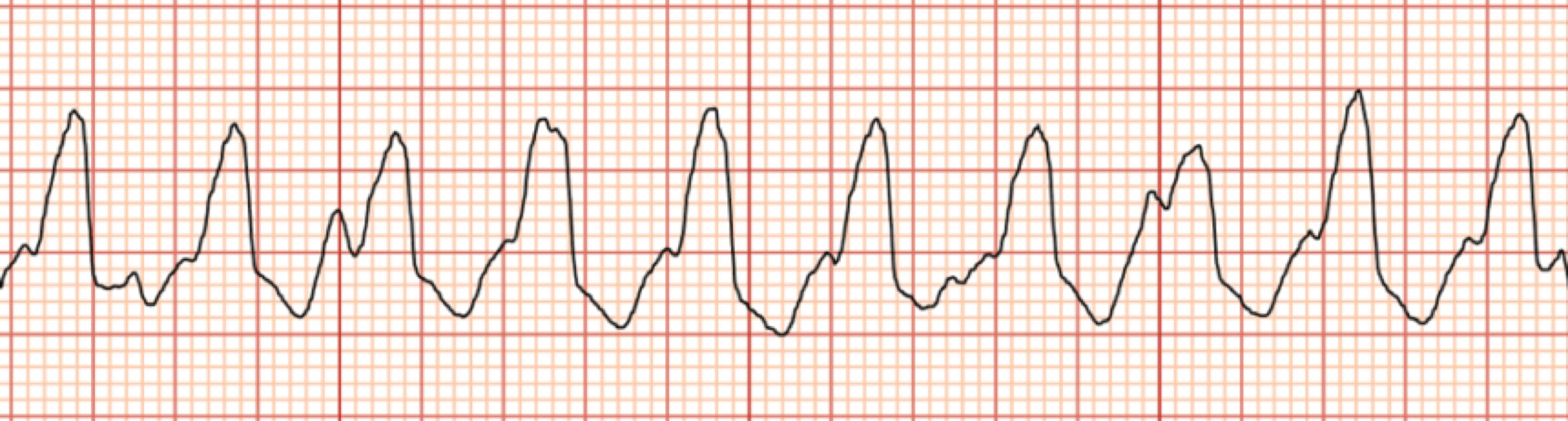
Agonal Rhythm:
Agonal rhythm: results when all the pacemakers of the heart (SA node, AV node, Purkinje fibers) have failed.
The tracing shows a wide, unusual QRS complex with no P or T wave.
The ventricular rate is less than 20/min.
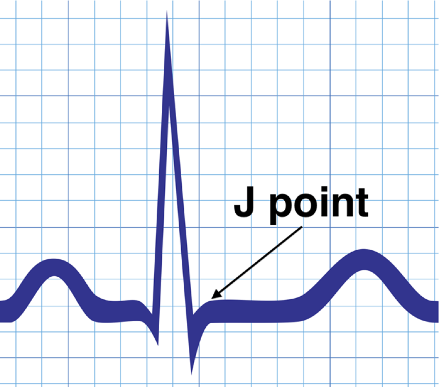
The J Point:
The J point represents the exact point in time where ventricular depolarization stops and ventricular repolarization starts.
The J point occurs at the end of the QRS complex, or where the ST segment begins.
During myocardial ischemia the J point can elevate or depress below baseline.
In terms of an EKG, think of acute myocardial injury and infarction as a problem of repolarization. This will prompt you to look for anomalies on the right side of the QRS complex.
Ischemia:
The classic pattern of myocardial ischemia is ST segment depression and/or elevation. Deeply inverted T waves are frequently encountered presentation of ischemia.
Injury:
The classic pattern of myocardial injury is ST segment elevation myocardial infarction (STEMI).
The Many Faces of Ischemia:
The tracings on the following strips represent common electrocardiographic findings suggestive of myocardial ischemia.
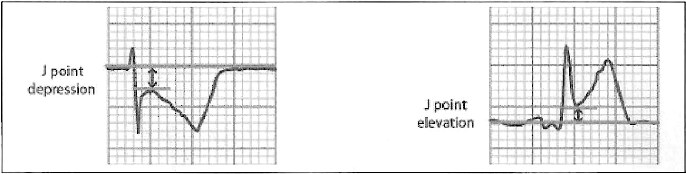
The "J" Point
If the J point is depressed or elevated, as shown on the tracings that follow, it can suggest ischemia.
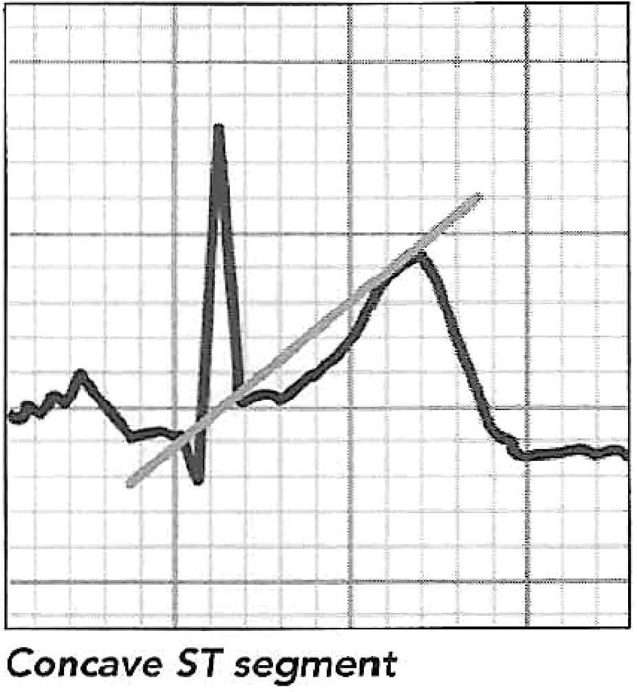
Concave:
A concave ST segment favors benign conditions.
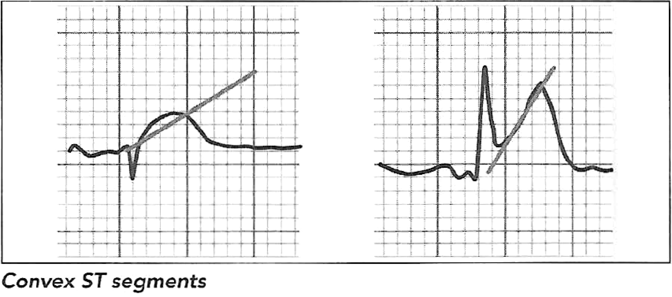
Convex:
A convex ST segment favors ischemia.
Test for this by drawing a line from the J point to the peak of the T wave, as shown below. If the line superimposes or if the T wave is above the line, the segment is convex.
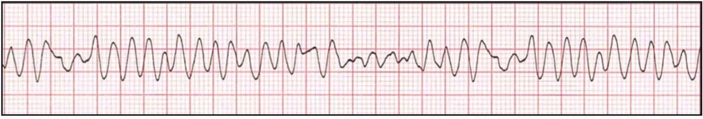
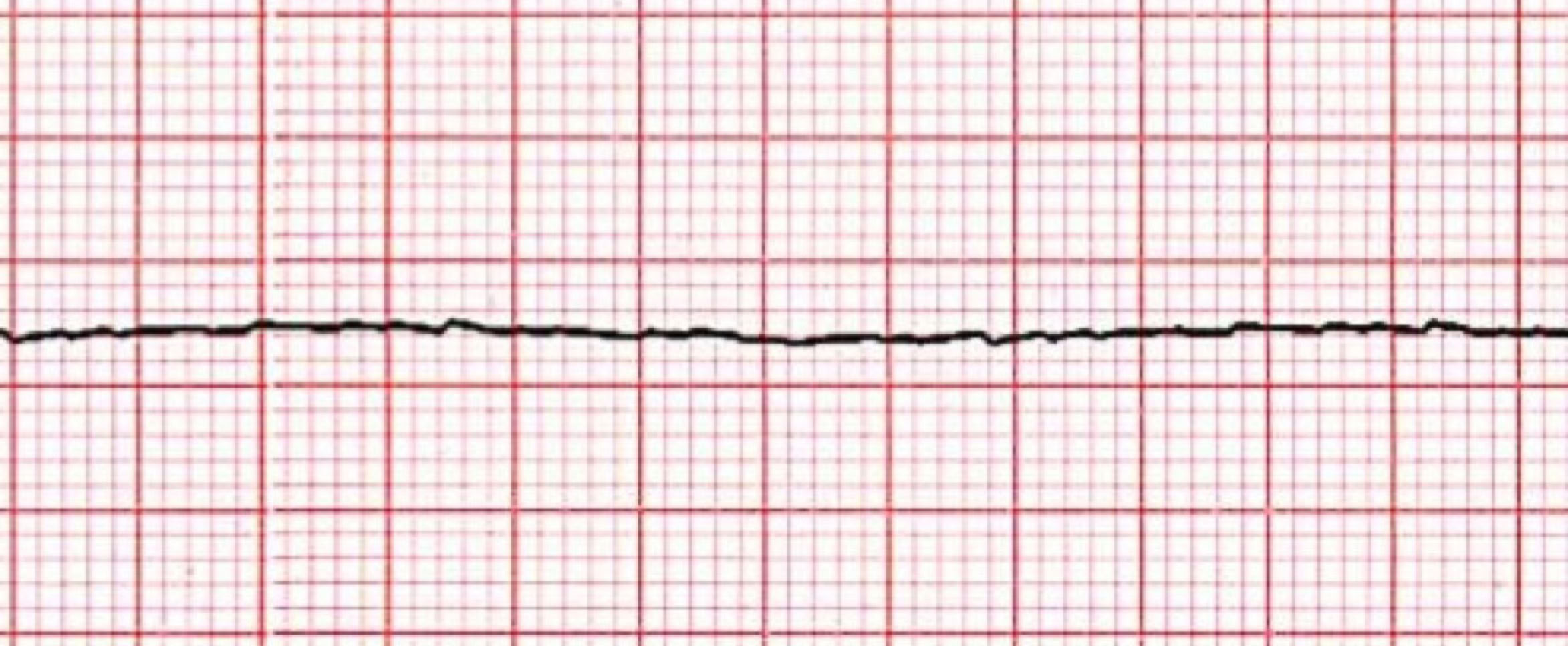
Asystole:
Asystole is the complete cessation of electrical activity in the heart.
Common causes of asystole include large pulmonary embolism, large myocardial infarction, respiratory arrest (hypoxia) and overdose.
If the technician recognizes asystole on the EKG, another lead should immediately be checked to confirm asystole.
If a patient's rhythm deteriorates to asystole, the technician should immediately call for help using the established guidelines for the facility.
The technician should initiate CPR as quickly as possible, ensure emergency services are notified, and send another person to retrieve the AED, if available.
If the patient does not positively react to the intervention, death will inevitably occur.