This chapter provides the information necessary to systematically approach the EKG and interpret arrhythmias encountered in the clinical setting. In addition, the chapter includes pictures and real EKG tracings to promote rhythm recognition. Important terms are located at the end of the chapter in a special section, enabling students to locate definitions quickly. The chapter concludes with a 10-question self-assessment drill. Calculate a Patient's Heart Rate from the EKG Tracing Heart rate can be measured from the EKG using a variety of different approaches. The following are the most commonly employed methods.
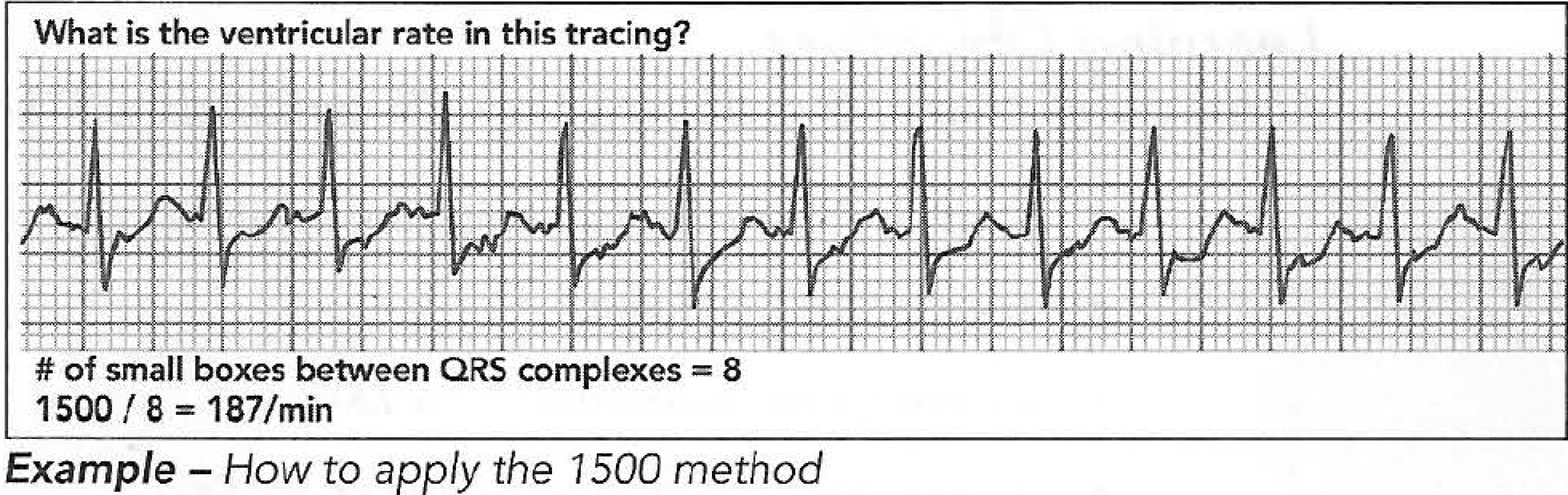
The 1500 method - Calculate the heart rate using the 1500 method by counting the number of small boxes between the P-P interval (for atrial rate) or R-R interval (for ventricular rate), then dividing 1,500 by that number. Fifteen hundred represents the number of small boxes, or the number of mm of paper consumed in one minute of time at the standard 25 mm/second paper speed. For example, if there are 15 small boxes between two R waves (R-R interval), the heart rate equals 1500/15, or 100/min. This is a great method for very precise measurements and is best applied to fast rhythms.
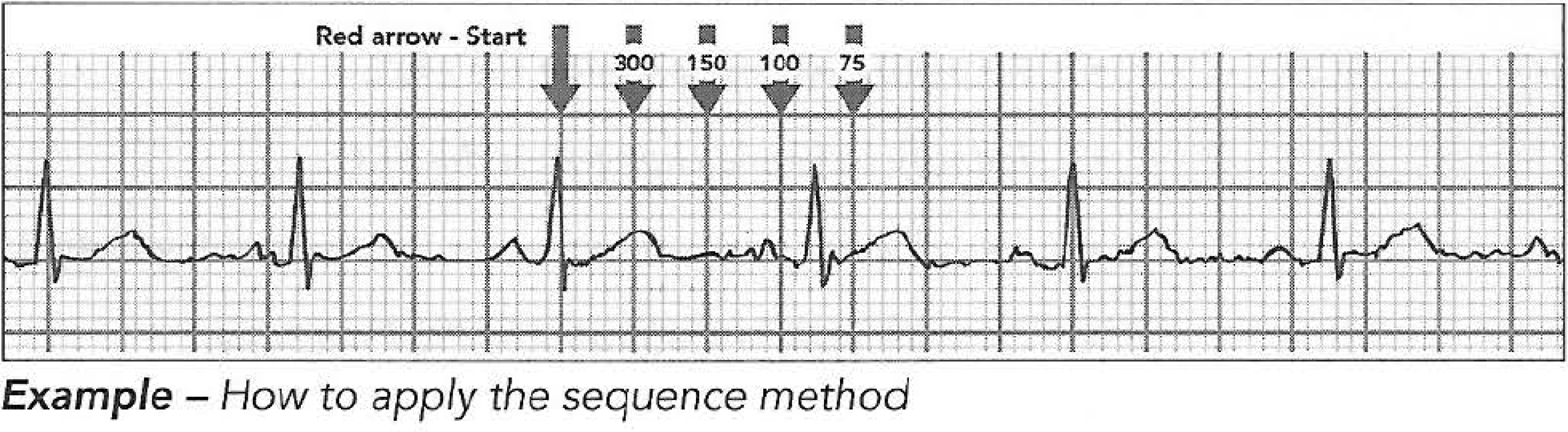
Sequence method -
The sequence method (also known as the 300 method) is derived
from the 1500 method. There are 300 large, 5 mm boxes in every minute of EKG tracing at the normal25 mm/second print speed. Remember the pattern 300-150-100-75-60-50. Calculate these numbers by dividing 300 by the number of large boxes between QRS complexes. Find an R wave and start counting away from it moving towards the right of the tracing in 5 mm segments (one large box). With every move, apply the number in the pattern you memorized. This is a wonderful rule to apply most of the time, with one
exception: irregular rhythms.
6-second rule -
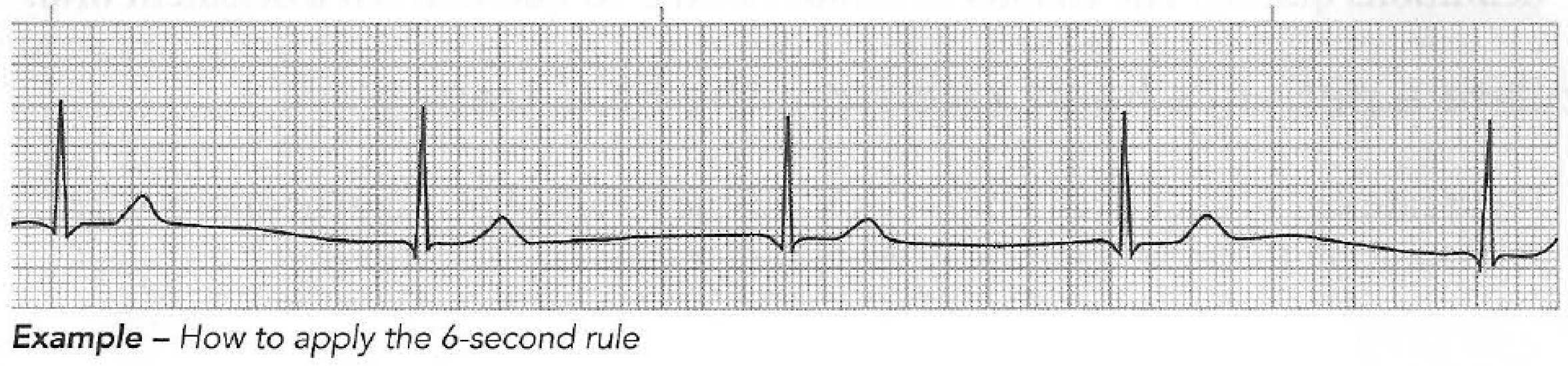
The 6-second rule is simple and works well in any situation. It is the gold standard for estimating the rate of an irregular rhythm. At the top of the tracing, there are small hash marks indicating 3-second intervals. Count the number of QRS complexes in two of the sections (6-second period) and multiply by 10. For example, you count six QRS complexes in 6 seconds. 6 X 10 = 60/min.
Identify artifacts from the tracing
Artifact can come from a variety of both patient and nonpatient factors. The most common reasons for artifact on the EKG caused by the patient include seizure, trembling, fast breathing, dry or wet skin, and shivering. Nonpatient causes include use of electrodes with dry gel, damaged patient cables, electromagnetic frequency, and interference from cell phones and other medical and nonmedical devices.
Resolve artifacts from the tracing
Wandering baseline - A wandering baseline often appears on the EKG when the electrodes are improperly placed on the patient's torso. The wandering baseline artifact can result from a variety of causes that include movement of the cables or leads during the reading, patient movement, loose electrodes, dry electrodes, a patient's labored breathing, or improper skin preparation, leaving traces of lotion, oils, or gel on the skin. An easy way to eliminate the wandering baseline is to move the limb leads to the wrists and ankles. Another way to reduce the artifact is to have the patient relax and breathe
more slowly, or to clean the skin where the electrodes are placed to remove lotion or oil, which causes a poor connection between the skin and the leads.
Seizure-
Seizure activity will cause huge artifact problems on the EKG. Seizure activity
must be controlled prior to acquiring the EKG tracing.
Trembling-
Some patients may be anxious, cold, or have an essential tremor. The EKG
technician must reassure the patient to keep him/her calm, provide warm blankets to control shivering, and attempt to move the electrodes to an area with minimal tremor.
Dry skin -
If the patient's skin is very dry, the electrodes and gel won't adhere well to
the skin. The gel will be unable to have the appropriate surface area contact to ensure a strong signal. The technician can reduce artifact from dry skin by gently abrading the skin and using tincture of benzoin to promote good adhesion and surface contact.
Wet skin-
Some patients will be diaphoretic during EKG acquisition. The technician
can wipe the patient off with a towel and apply tincture of benzoin to the patient's skin before placing the electrodes. The benzoin needs to completely dry prior to applying the EKG electrodes.
Cold patient -
If the patient is very cold, the patient may shiver, and in some cases, the
electrodes won't adhere to the skin. The technician should provide the patient with warm blankets. Some patients may be unable to stop shivering, and the EKG may have to be acquired with the artifact.
Dry gel-
The gel on the EKG electrodes is specially designed to interface with the
patient's skin. The gel is able to sense extremely low levels of energy and requires the full surface area to make contact with the patient. Electrodes with dry gel should not be used.
Cell phone interference-
Cell phone interference can cause lots of artifact on the EKG. It may appear as flutter or P waves on the tracing at a rate of 300/min. Though the cell phone artifact morphology is different than normal P wave or flutter wave
morphology, take special care to ensure the patient's cell phone is off or moved away from the patient during the procedure.
Medical device interference-
Medical and other electronic devices can interfere with the EKG tracing. Ensure that all unnecessary devices are moved away from the patient or turned off. Medical devices are often designed to minimize interference with
other devices, so the technician should consider nonmedical devices as the source of interference first.