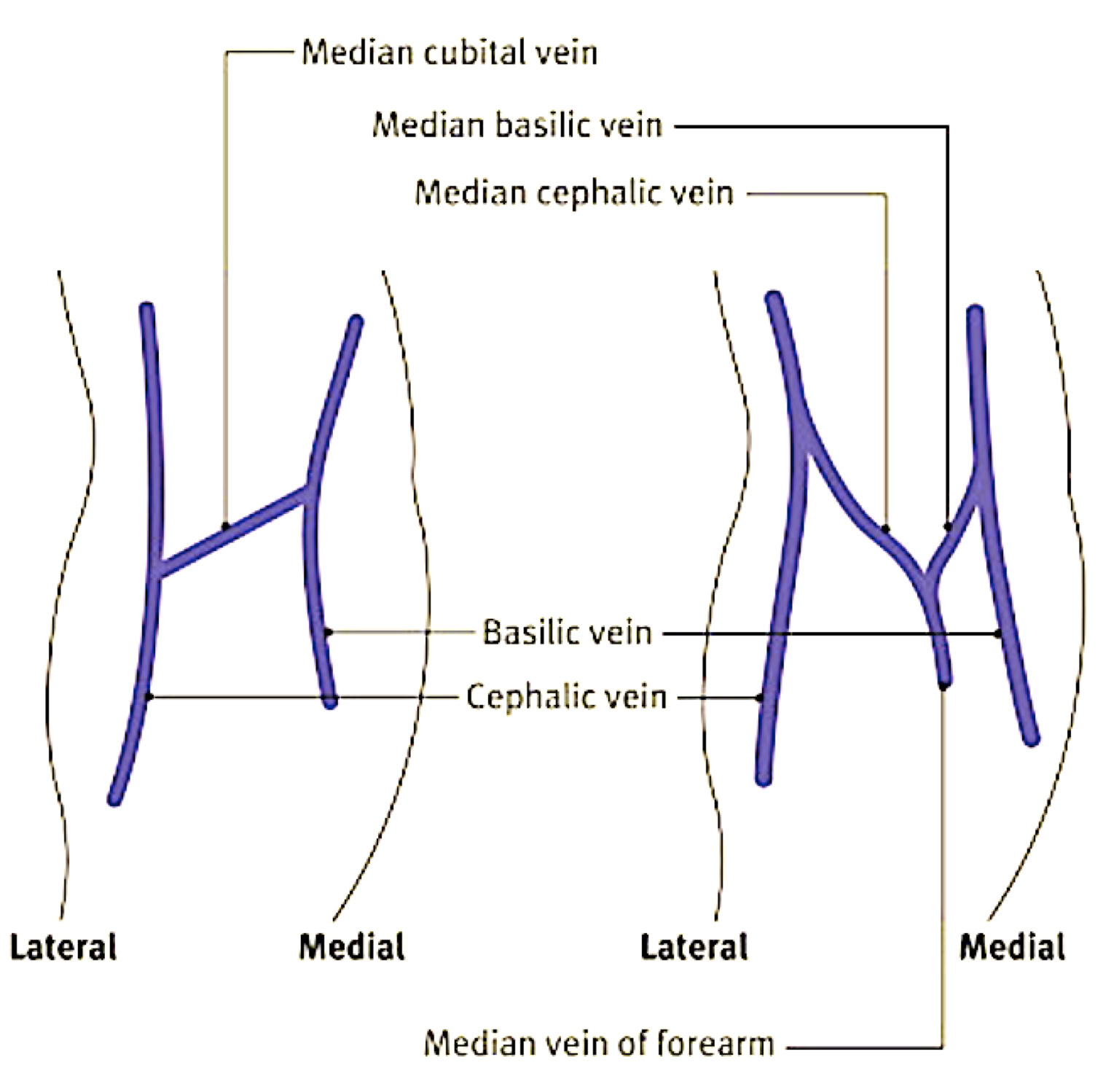
Shallow depression in arm anterior to & Below bend of elbow
Site of major veins, and thus first choice for venipuncture
H-shaped antecubital veins (in 70% of population )
Median cubital, cephalic, & basilic veins
M-shaped antecubital veins
Median cubital, cephalic, & median basilic veins
Veins on back of hand & wrist (less frequently used for venipuncture due to veins collapsing)
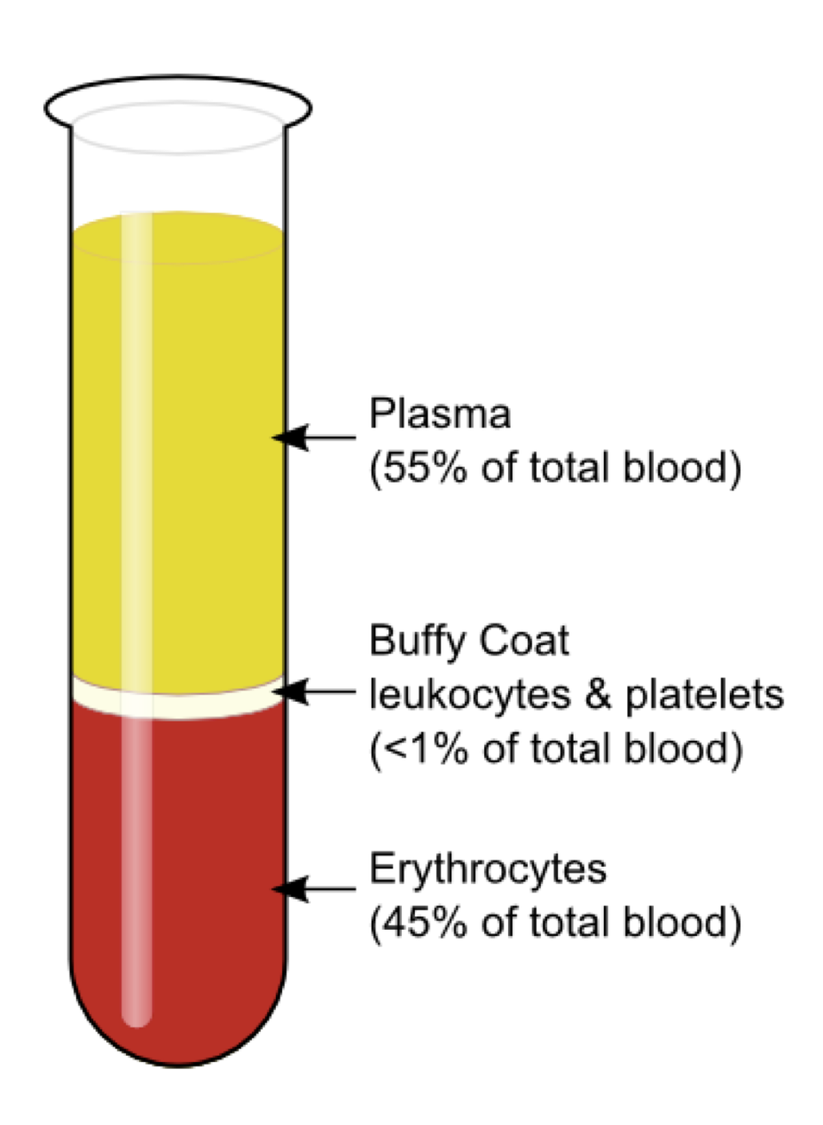
The Blood
An average adult has 5 to 6 Liters of blood. Blood is composed of plasma- the fluid portion-and cellular components, called the formed elements
Erythrocytes ( red blood cells )
RBCs carry hemoglobin, the iron-containing oxygen transport protein that gives blood its red color. A single RBC remains in the peripheral circulation
about 120 days before being removed by the liver.
Most numerous cells in the body
Carry oxygen and carbon dioxide
Produced in bone narrow
Leukocytes ( White blood cells )
WBCs, or leukocytes, protect the body against infection. WBCs are produced in the bone marrow and lymph nodes and undergo a complex maturation process.
Formed in bone marrow and lymphatic tissue
Neutralize or destroy pathogens
Plasma
Plasma constitutes 55% of the volume of blood. It is 90% fluid portion of whole blood separated from the RBCs WBCs & platelets
Contains fibrinogen
Serum
Fluid portion of blood remaining after clotting
Can be seperate from clot by centrifugation
Does not contain fibrinogen ( used for clotting)
-Notice: The “Buffy coat” is the white layer between the RBCs and plasma. It is made up of WBCs which FIGHT INFECTION.
Whole blood
Blood in the same form as it is in the
bloodstream
Hemostasis and Coagulation
Hemostasis refers to the processes by which blood vessels are repaired after injury. It occurs in a series of steps, from muscular contraction of the vessel walls, through clot formation, to removal of the clot when the vessel repairs itself.
Hemostasis is the arrest or stoppage of bleeding after injury as a body response
Requires coordinated interaction of endothelial cells lining blood vessels, platelets, other blood cells, plasma proteins & clotting
Vessel Healing Process (4 interrelated responses )
1.) vasoconstriction
2.) formation of primary platelet plug
3.) progression to a stable blood clot
4.) fibrinolysis (dissolving of clot)
1.) Vasoconstriction
Rupture of a vein or artery causes an immediate vascular spasm, or contraction of the smooth muscle lining the vessel. This reduces the vessel diameter, substantially reducing the blood loss that would otherwise
occur. This contraction lasts about 30 minutes. For capillaries, this may be enough to allow the wound to seal.
2.) Formation of primary platelet plug
Exposure of materials beneath the endothelial lining causes platelets to stick to the endothelial cells almost immediately, a process known as adhesion.
Additional platelets then stick to these, a process known as aggregation.
3.) Progression to a stable blood clot
Coagulation involves a complex interaction of enzymes and other factors whose activation ultimately results in formation of a blood clot. A meshwork of fibrin, platelets, and other blood cells that closes off the wound. The coagulation cascade begins from 30 seconds to several minutes
after the injury.
4.) Fibrinolysis (dissolving of clot)
As the wound is closed and tissue repair commences, fibrin itself is broken down slowly, a process called fibrinolysis.