Carries carbon dioxide and other waste away from cells to excretory organs, kidney, lungs and skin
Aids in coagulation process
Assists in defending body against disease
And in regulation of body temperature
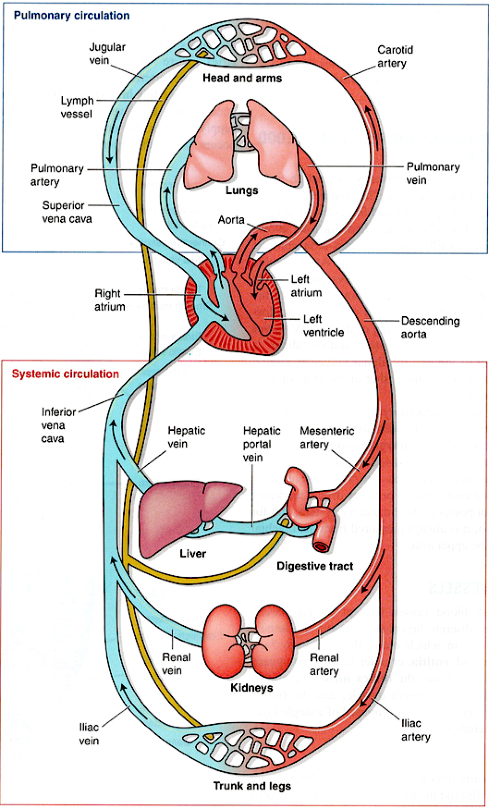
The circulatory system is a system of closed tubes. Circulation occurs in two large loops- the pulmonary circulation and the systemic circulation. The pulmonary circulation carries blood between the heart and the lungs for gas exchange, and the systemic circulation carries blood between the heart and the rest of the body's tissues. In both cases, arteries carry blood from the heart to capillary beds, where exchange occurs. Veins return blood to the heart.
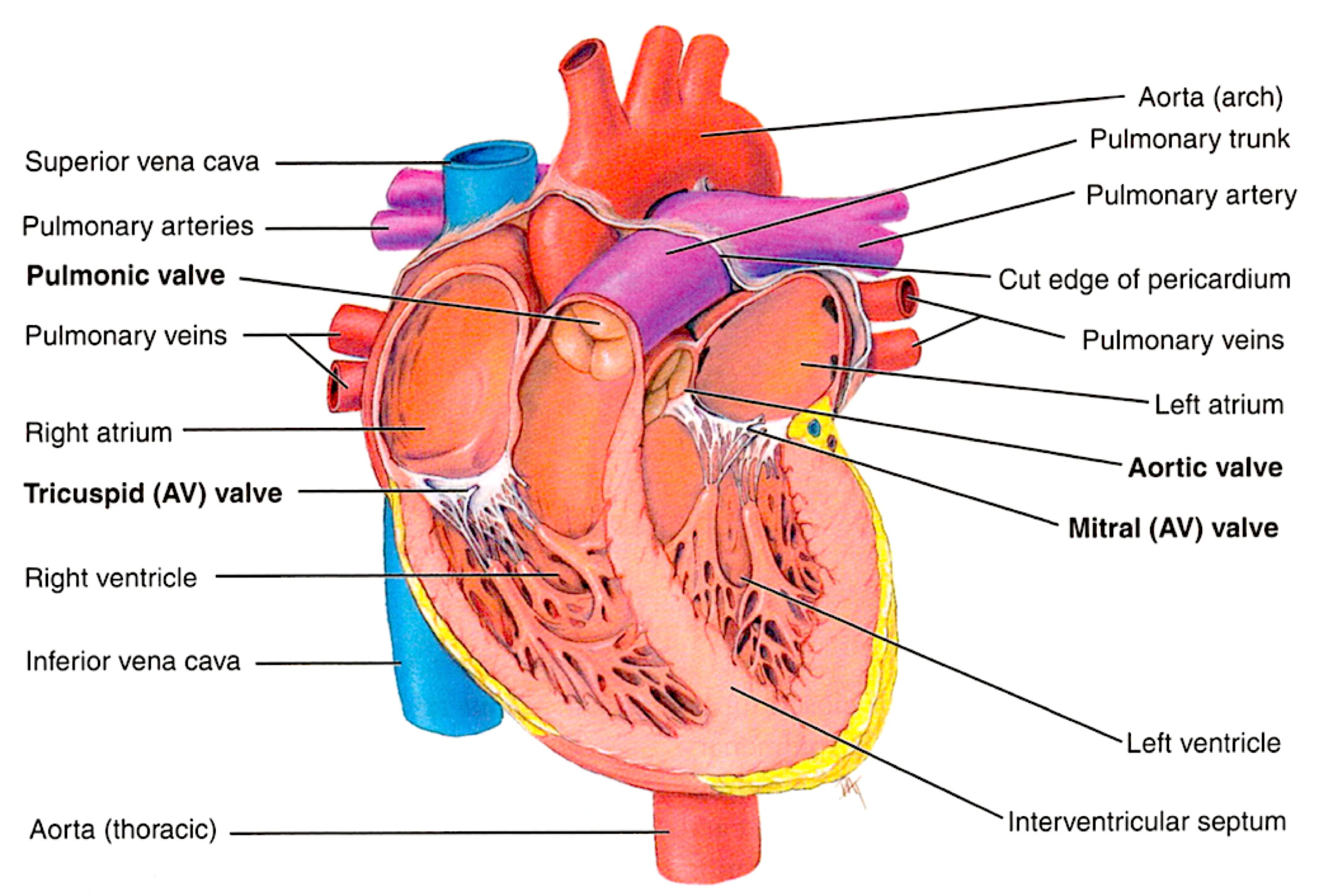
The Heart
4 chambers
Atria, right and left
Ventricles, right and left
Septa: partitions separating the right and left sides of the heart
Ischemia: inadequate supply of the oxygen to tissue, often caused by partial obstruction coronary artery
Myocardial infarction: heart attack, cause my complete obstruction of coronary artery
Cardiac cycle
One complete contraction (systole ) and relaxation ( diastole ) of the heart
Last about 0.8 seconds
Pulmonary circulation
Carries blood from right ventricle of heart to lungs
Carbon dioxide is removed: oxygen is picked up
Oxygenated blood returns to the left atrium of the heart
Systemic circulation
Carries oxygenated blood and nutrients from left ventricle to body cells
Returns deoxygenated blood with carbon dioxide and wastes from cells to right atrium
-Origin of the heart sounds
The first sound: “LUBB” (ventricles contract, AV valves close )
The second sound: “DUPP” ( ventricles relax, semilunar valves close )
Cardiac output
Heart rate: number of heartbeats per minute
Cardiac output: volume of blood pumped by the heart in one minute
Pulse: palpable rhythmic dropping caused by alternating expansion and contraction of an artery as blood passes through
Blood pressure: force exerted by blood on the walls of vessels
Arteries
Carry oxygenated blood away from the heart to issue
Thick walls to withstand high pressure
Aorta is the largest artery
Veins
Return deoxygenated blood from tissue to heart
Thin walls, low pressure
Blood is moved by skeletal muscles movement, valves that prevent back flow, & pressure changes in breathing
Capillaries
Microscopic, one-cell-thick vessels that connect arterioles & veins
Site of gas exchange between blood and body tissues
Internal space a blood vessel through which blood flows
Valves
-thin membrane leaflets in veins prevents back flow of blood
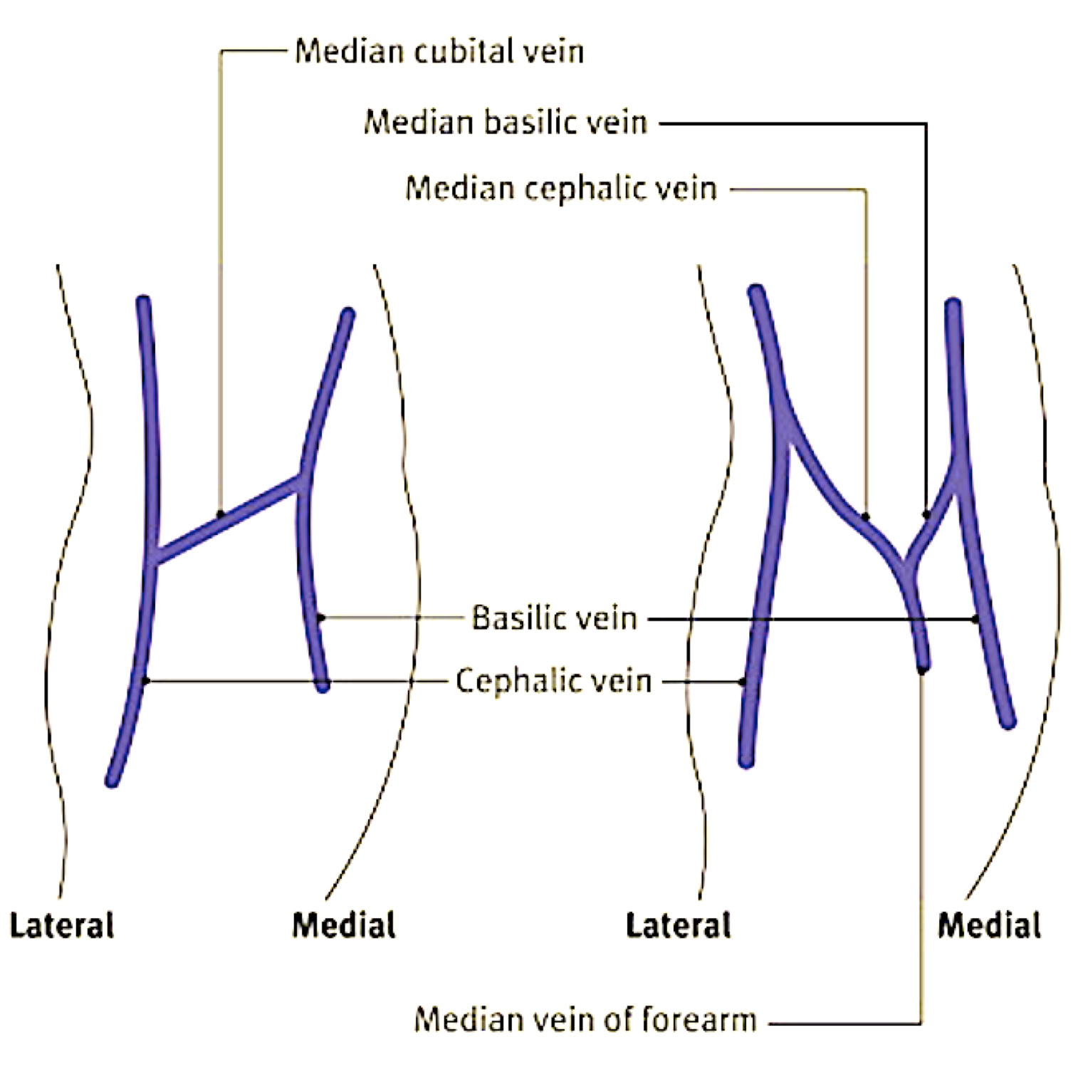
Phlebotomy related vascular anatomy
Antecubital fossa:
Shallow depression in arm anterior to & Below bend of elbow
Site of major veins, and thus first choice for venipuncture
H-shaped antecubital veins (in 70% of population )
Median cubital, cephalic, & basilic veins
M-shaped antecubital veins
Median cubital, cephalic, & median basilic veins
Veins on back of hand & wrist (less frequently used for venipuncture due to veins collapsing)
The Blood
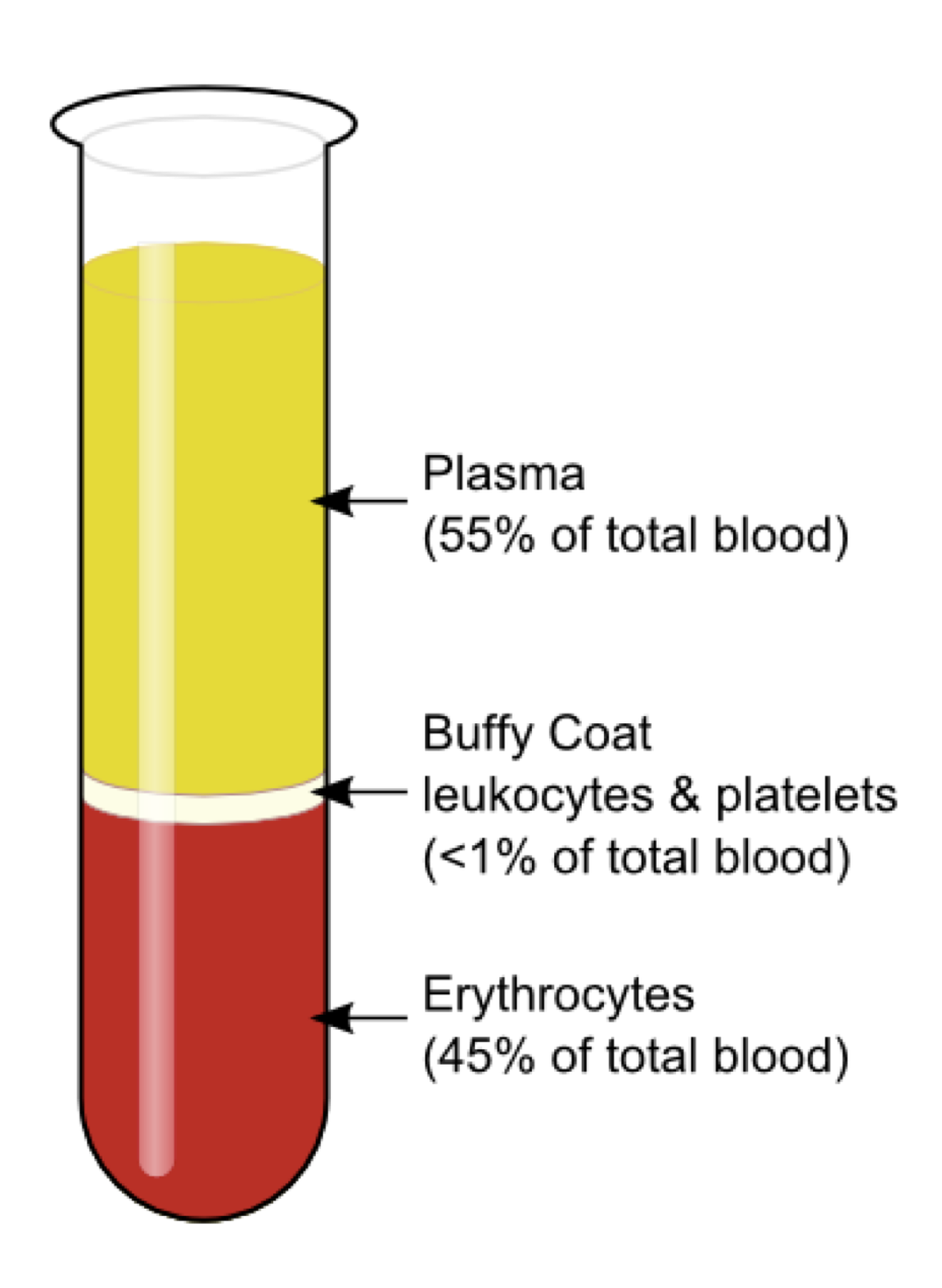
An average adult has 5 to 6 L of blood. Blood is composed of plasma- the fluid portion-and cellular components, called the formed elements
Erythrocytes ( red blood cells )
RBCs carry hemoglobin, the iron-containing oxygen transport protein that gives blood its red color. A single RBC remains in the peripheral circulation
about 120 days before being removed by the liver, bone marrow, or spleen.
Most numerous cells in the body
Carry oxygen and carbon dioxide
Produced in bone narrow
Leukocytes ( White blood cells )
WBCs, or leukocytes, protect the body against infection. WBCs are produced in the bone marrow and lymph nodes and undergo a complex maturation process.
Formed in bone marrow and lymphatic tissue
Neutralize or destroy pathogens
Plasma
Plasma constitutes 55% of the volume of blood. It is 90% fluid portion of whole blood separated from the RBCs WBCs & platelets
Contains fibrinogen
Serum
Fluid portion of blood remaining after clotting
Can be seperate from clot by centrifugation
Does not contain fibrinogen ( used for clotting)
-Notice: The “Buffy coat” is the white layer between the RBCs and plasma. It is made up of WBCs which FIGHT INFECTION.
Whole blood
Blood in the same form as it is in the
bloodstream
Not allowed to clots or separate
Specimen must be connected in anticoagulant tube
Must be mixed a minimum of two minutes just prior to test
Hemostasis and Coagulation
Hemostasis refers to the processes by which blood vessels are repaired after injury. It occurs in a series of steps, from muscular contraction of the vessel walls, through clot formation, to removal of the clot when the vessel repairs itself.
Arrest or stoppage of bleeding after injury as a body response
Requires coordinated interaction of endothelial cells lining blood vessels, platelets, other blood cells, plasma proteins & clotting
Process (4 interrelated responses )
1.) vasoconstriction
2.) formation of primary platelet plug
3.) progression to a stable blood clot
4.) fibrinolysis (dissolving of clot)
1.) Vasoconstriction
Rupture of a vein or artery causes an immediate vascular spasm, or contraction of the smooth muscle lining the vessel. This reduces the vessel diameter, substantially reducing the blood loss that would otherwise
occur. This contraction lasts about 30 minutes. For capillaries, this may be enough to allow the wound to seal.
2.) Formation of primary platelet plug
Exposure of materials beneath the endothelial lining causes platelets to stick to the endothelial cells almost immediately, a process known as adhesion.
Additional platelets then stick to these, a process known as aggregation.
3.) Progression to a stable blood clot
Coagulation involves a complex interaction of enzymes and other factors whose activation ultimately results in formation of a blood clot. A meshwork of fibrin, platelets, and other blood cells that closes off the wound. The coagulation cascade begins from 30 seconds to several minutes
after the injury.
4.) Fibrinolysis (dissolving of clot)
As the wound is closed and tissue repair commences, fibrin itself is broken down slowly, a process called fibrinolysis.
Lorem ipsum dolor sit amet, consectetur adipiscing elit. Aliquam tincidunt lorem enim, eget fringilla turpis congue vitae. Phasellus aliquam nisi ut lorem vestibulum eleifend. Nulla ut arcu non nisi congue venenatis vitae ut ante. Nam iaculis sem nec ultrices dapibus. Phasellus eu ultrices turpis. Vivamus non mollis lacus, non ullamcorper nisl. Pellentesque habitant morbi tristique senectus et netus et malesuada fames ac turpis egestas. Phasellus sit amet scelerisque ipsum. Morbi nulla dolor, adipiscing non convallis rhoncus, ornare sed risus. Sed adipiscing eget nibh at convallis. Curabitur eu gravida mauris, sit amet dictum metus. Sed a elementum arcu. Proin consectetur eros vitae odio sagittis, vitae dignissim justo sollicitudin. Phasellus non varius