Anatomy and Physiology / Introduction to the Heart
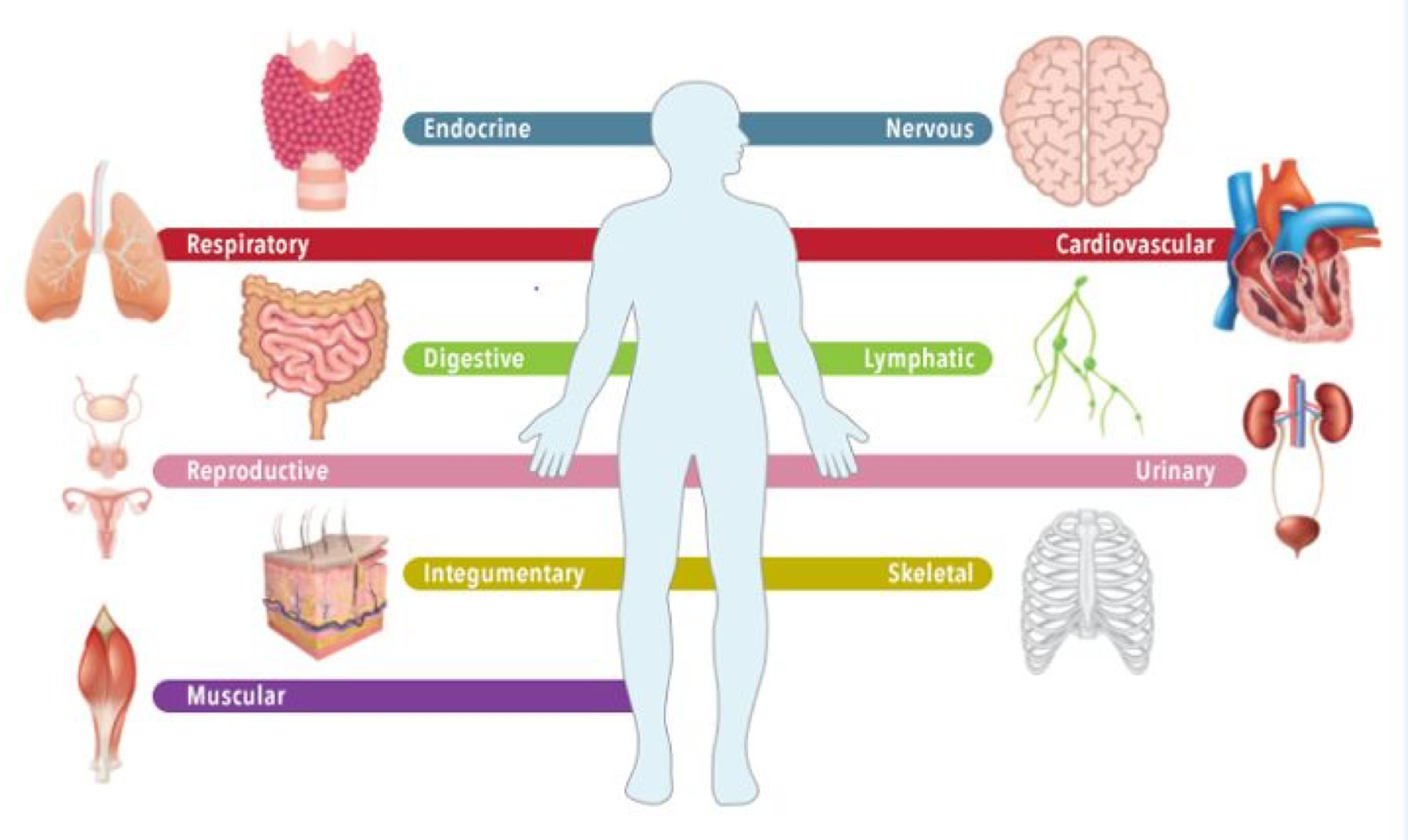
The human body is broken down into 11 systems. These systems are often described in terms of Anatomy (the study of parts of the body systems), and Physiology (or the study of how the body system functions).
These body systems have specific functions that work together to maintain homeostasis.
Homeostasis:
A state of equilibrium or balance in the body internal environment maintained by compensating for charges ( feedback and regulation )
Anatomical terms we will be using are:
Axilla: armpit
Thorax: chest
Thoracic cavity: area inside the chest
Mediastinum: central compartment of the thoracic cavity where heart is located.
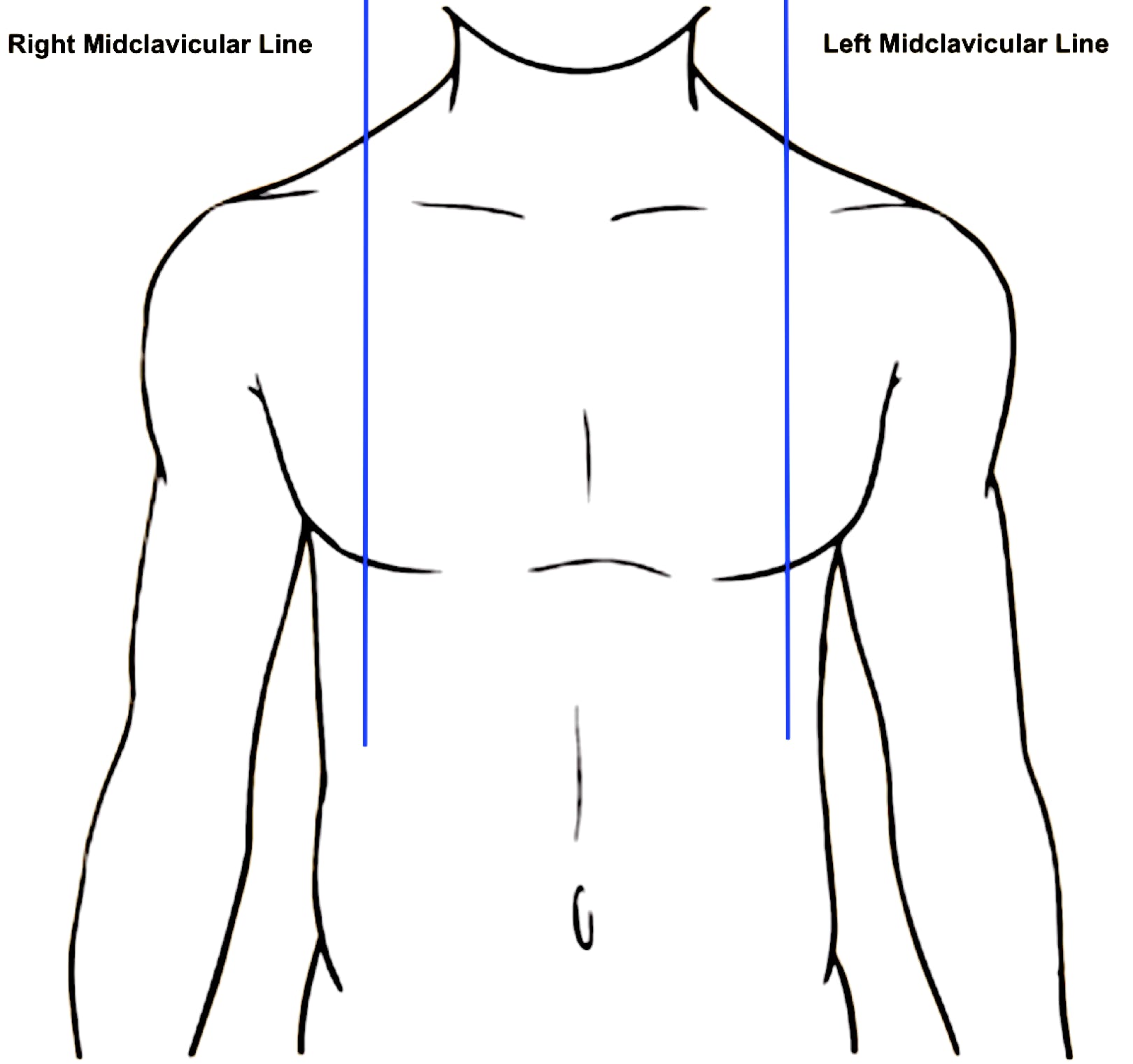
Clavicle: collarbone
Sternum: breastbone.
Midclavicular line: imaginary line parallel to the midline and passing through the midpoint of the clavicle on the anterior surface of the body.
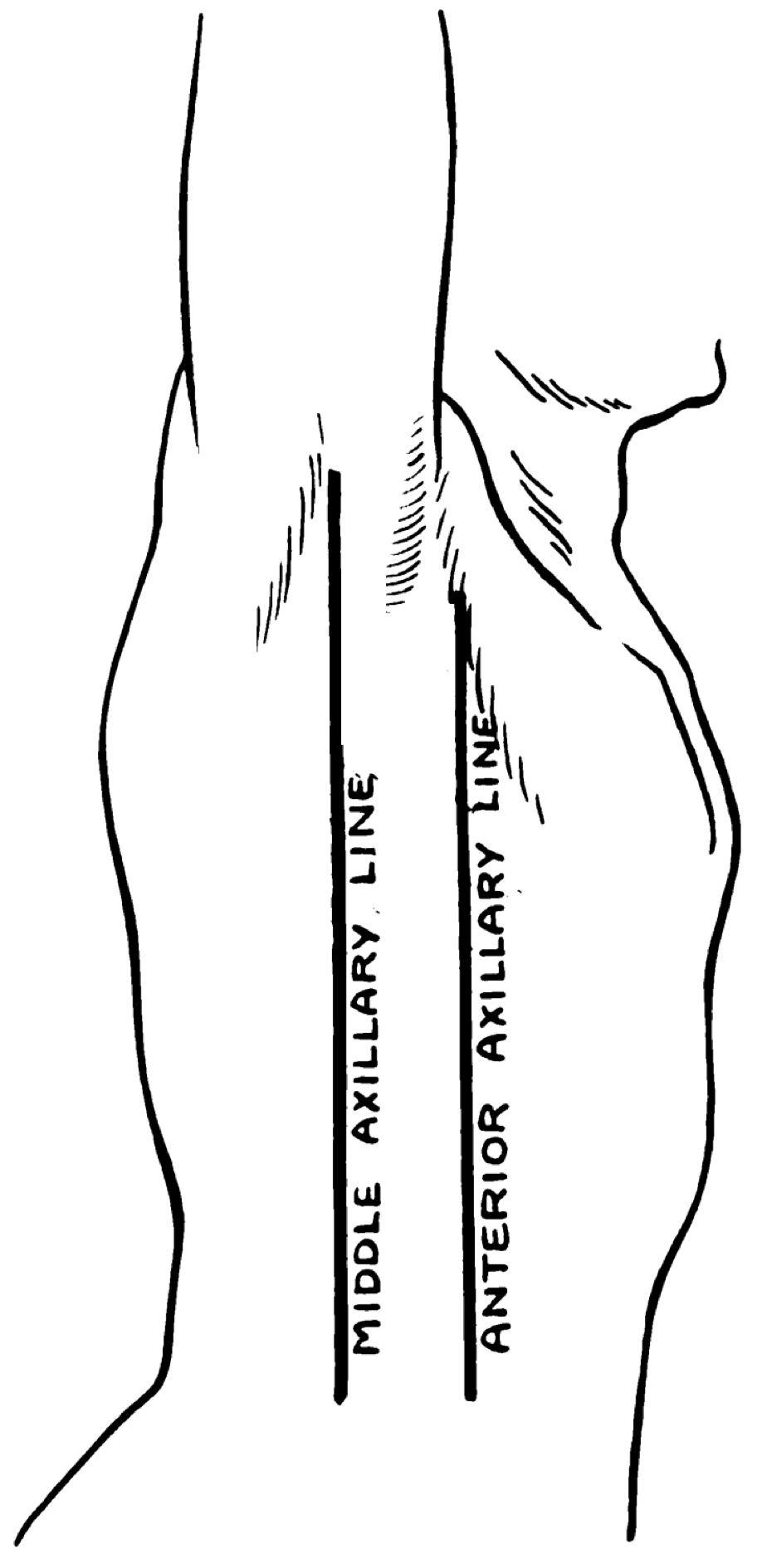
Anterior axillary line: imaginary line parallel to the midclavicular line beginning at the anterior axillary fold.
Midaxillary line: imaginary line parallel to the anterior axillary line beginning at the midpoint of the axillary fold under the arm.
The Human Heart
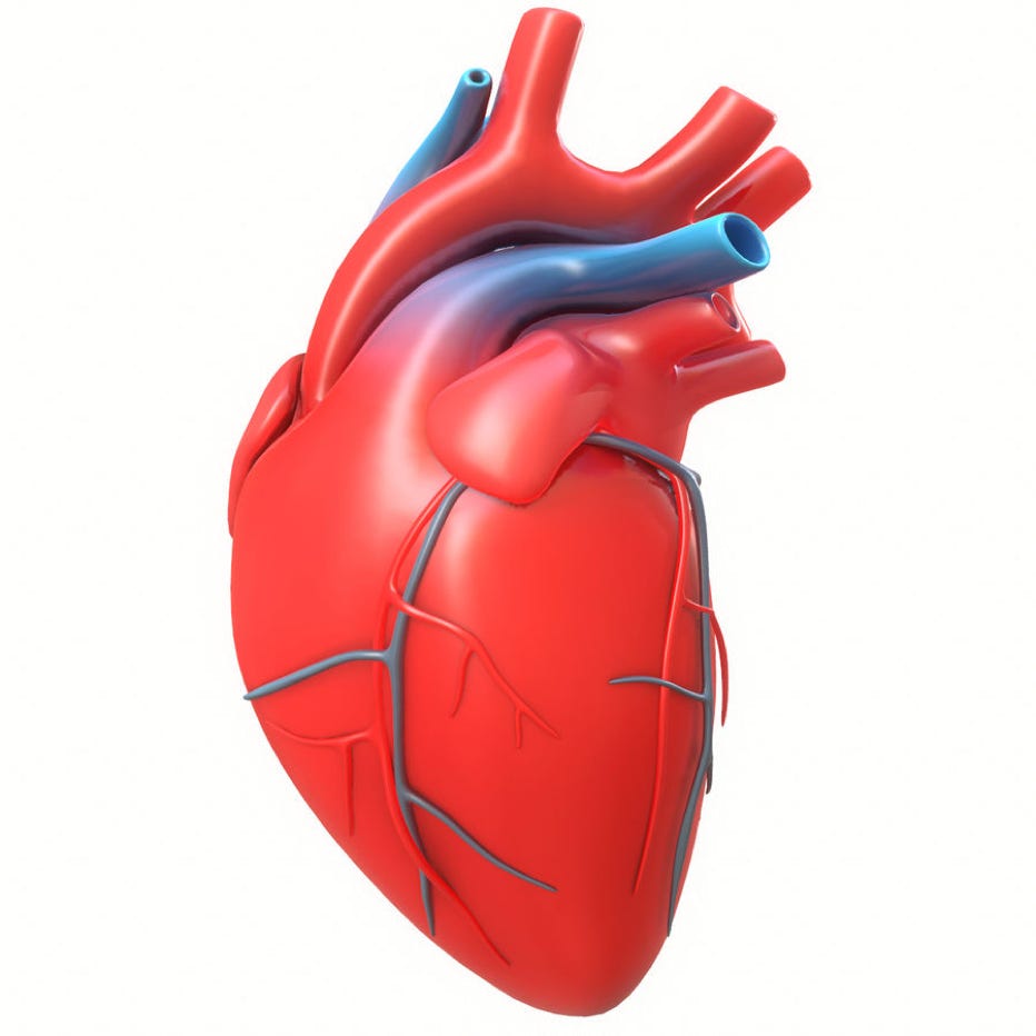
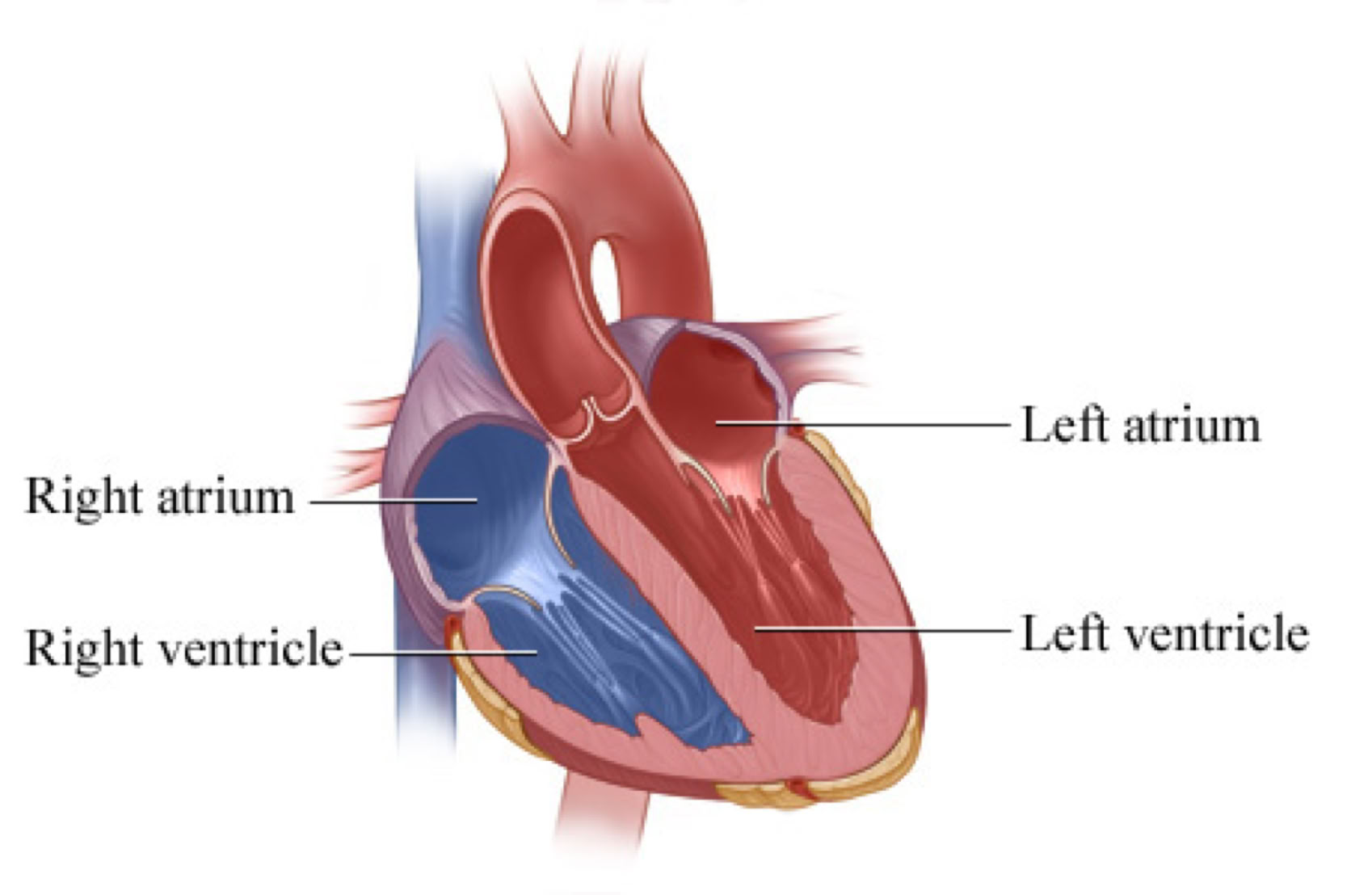
The human heart is a four-chambered organ responsible for supplying oxygenated blood to the entire body.
The four chambers consist of the right atrium and left atrium (plural of atrium is atria), and the right and left ventricles.
The right and left sides of the heart are separated by a wall called septum.
The heart has two upper chamber called atria which receive blood which is then passed down to the ventricles.
The two lower chambers are called ventricles which either pump blood towards the lungs for oxygenation or to the body for perfusion.
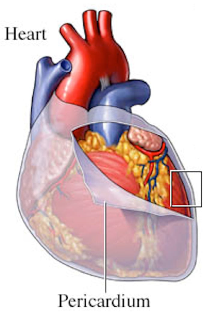
The heart wall consists of three distinct layers: the epicardium, myocardium, and endocardium.
The epicardium, or outermost layer, is made of connective tissue that forms a sac around the heart, known as the pericardium.
The pericardial sac completely surrounds the heart and provides protection and lubrication between the heart and other organs in the chest.
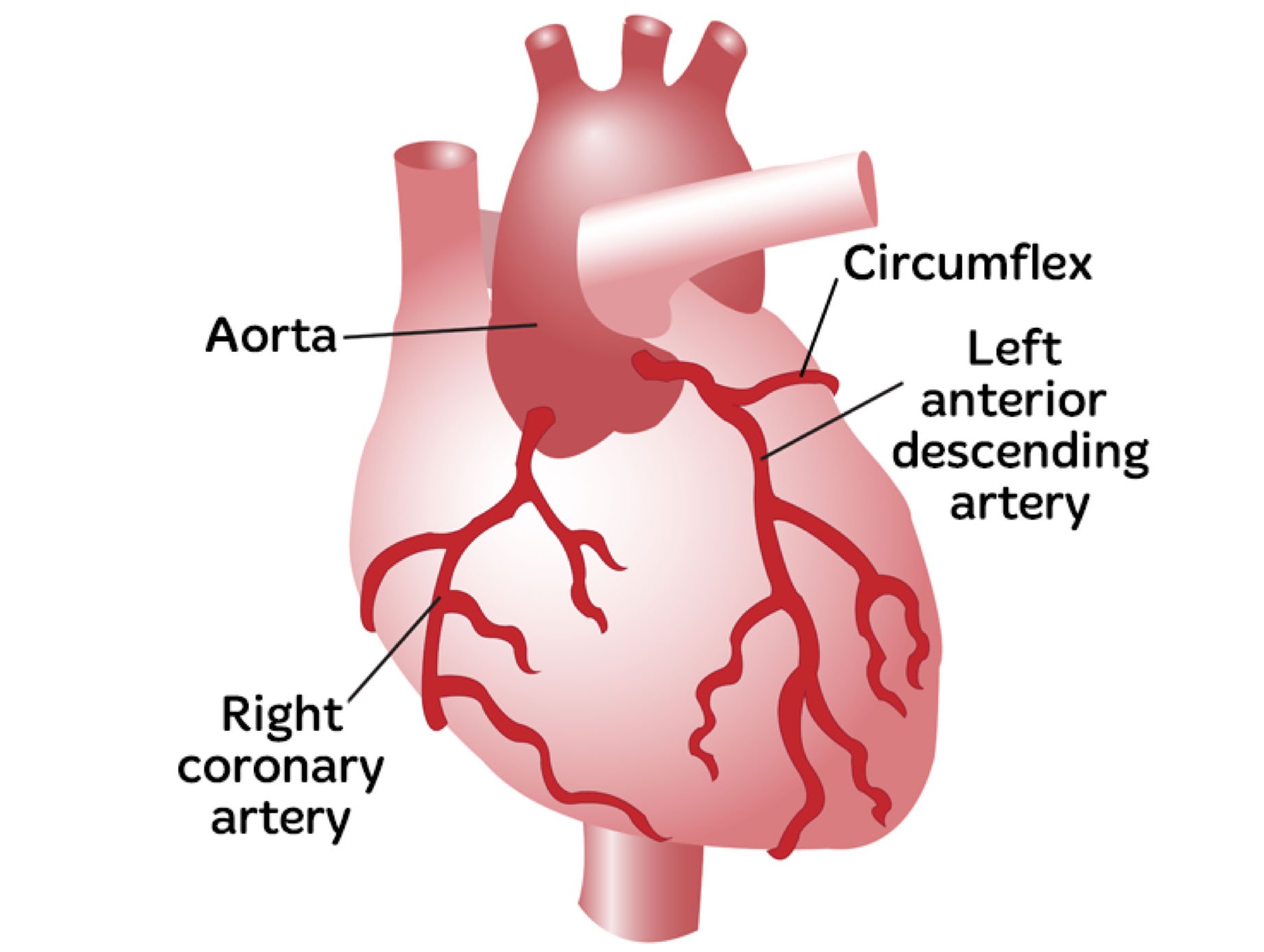
Right and Left Coronary Arteries:
The coronary arteries carry oxygenated blood to the atria and ventricles.
Right coronary artery (RCA) provide blood to the right atrium, right ventricle, the Sinoatrial node and Atrioventricular node.
Left coronary artery (LCA) carries oxygenated blood to the myocardium and bifurcates into the left circumflex artery (LCX), and the left anterior descending artery (Lad).
The left circumflex artery supplies oxygenated blood to the posteriolateral aspect of the left ventricle.
The left anterior descending artery supplies the anterior wall of the left ventricle. Left anterior occlusion can lead to ventricle arrhythmias and death.
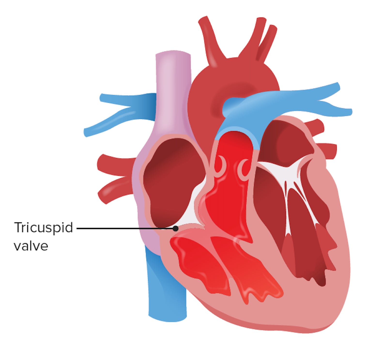
A series of valves ensures that blood flows in the correct direction as it is pumped through the heart. The valves of the heart are made up of endocardium tissue.
The four major valves of the heart are.
Tricuspid valve
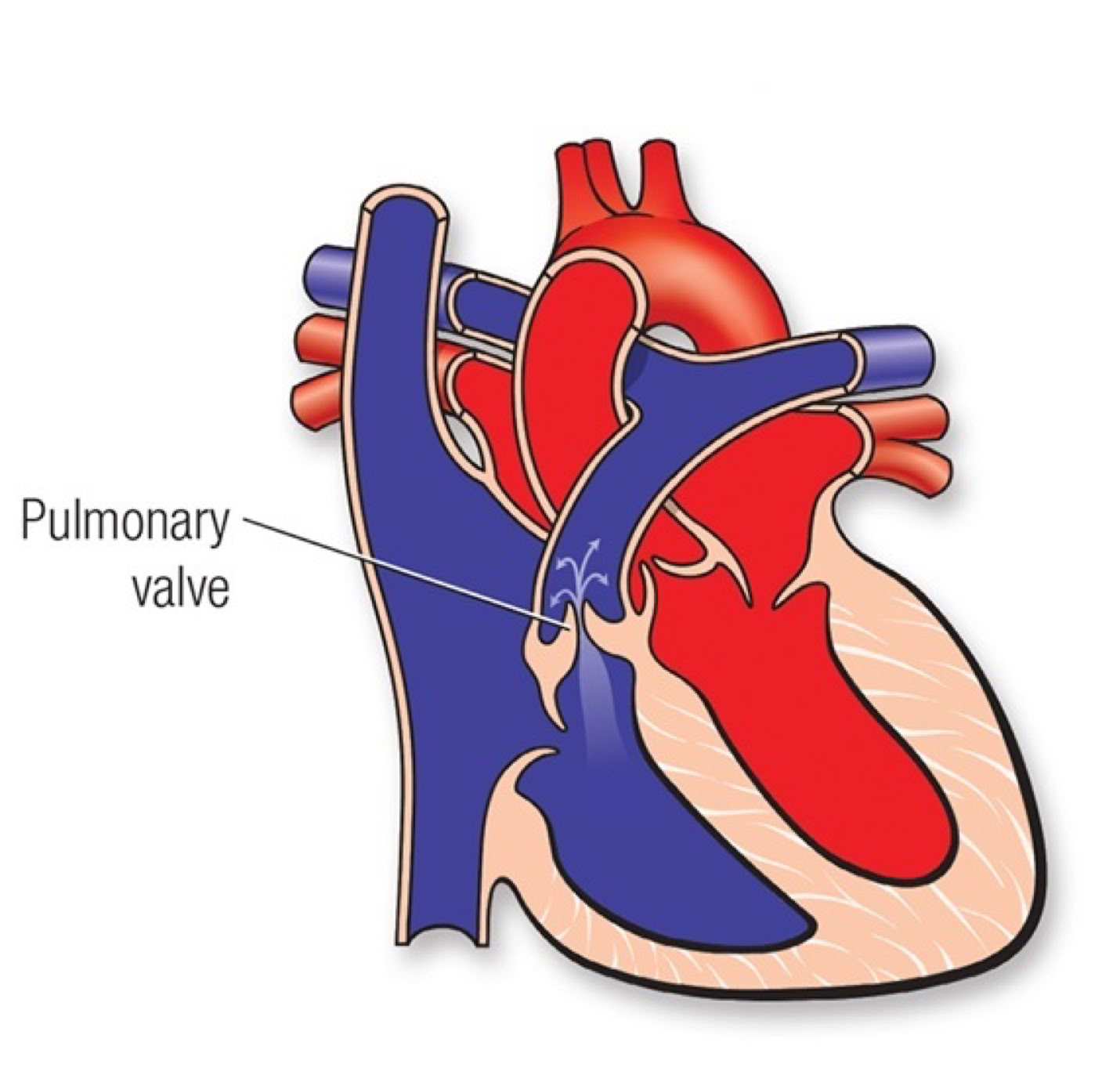
Pulmonary valve
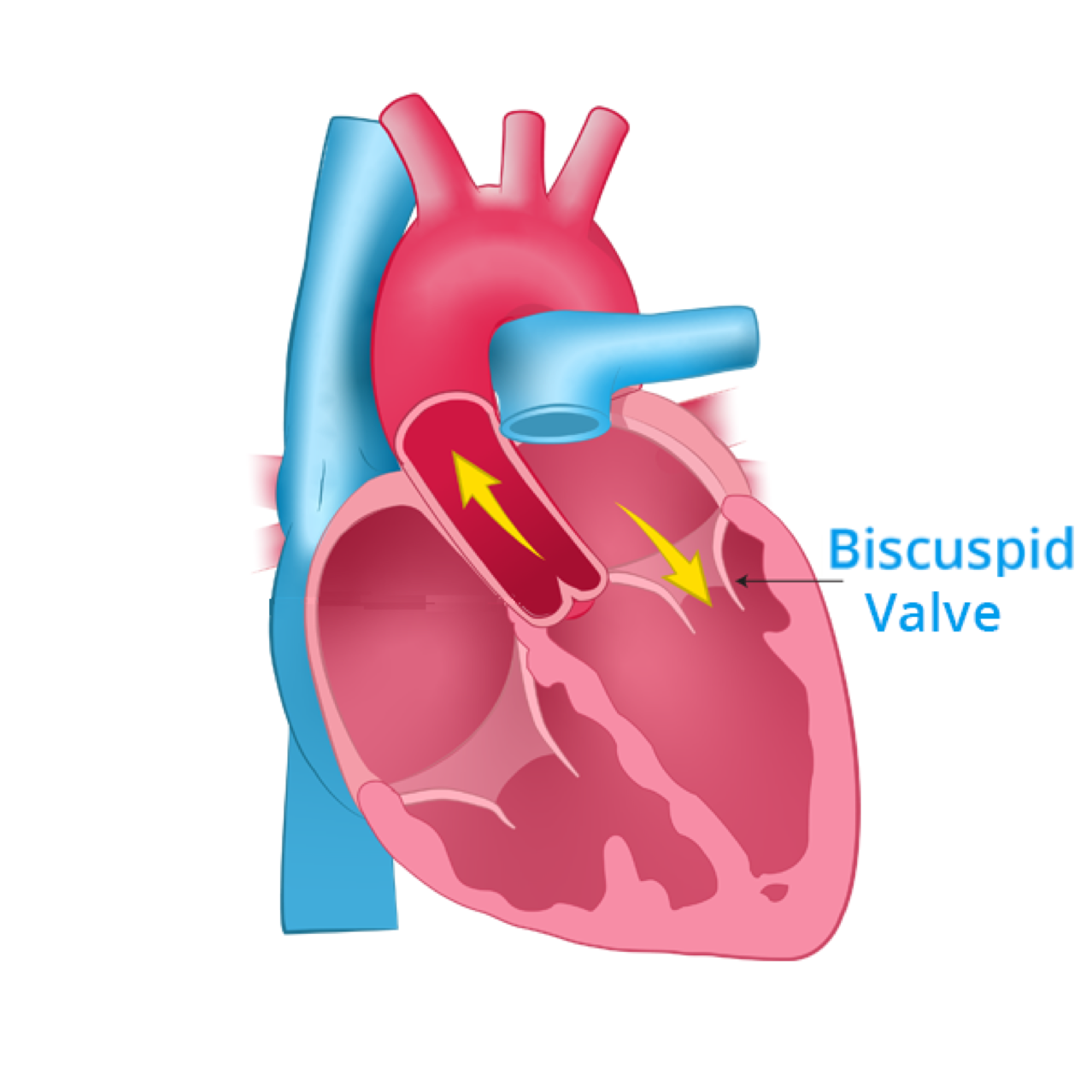
Bicuspid valve (mitral)
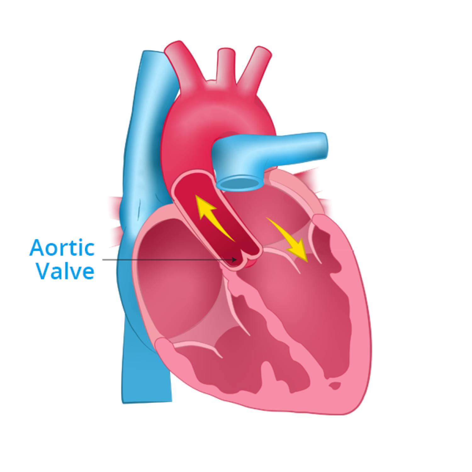
Aortic valve
Tricuspid valve: (also called the right atrioventricular valve), is found between the right atrium and the right ventricle
It is made up of three flaps (or cusps) connected to special tissues in the ventricular wall.
The function of the tricuspid valve is preventing the backward flow of the blood when the right ventricle contracts.
Pulmonary valve: ( pulmonary and aortic valve are also called semilunar valve) is located between the right ventricle and the pulmonary artery. This valve has three cusps that are shaped like pockets.
These pockets fill and close the valve as the pulmonary artery fills and pressure in the pulmonary artery increases.
Bicuspid valve: (also called the mitral valve) is between the left atrium ant the left ventricle. Is in the left side of the heart. This valve has two cusps that are connected to special tissues into the ventricular wall. These cusps prevent the backward flow of blood during left ventricular contraction.
Aortic valve: (also called the aortic semilunar valve) is located between the left ventricle and the aorta.
This valve consists of three cusps, or flaps.
The closed aortic valve keeps blood from flowing back into the heart, directing blood to the body.
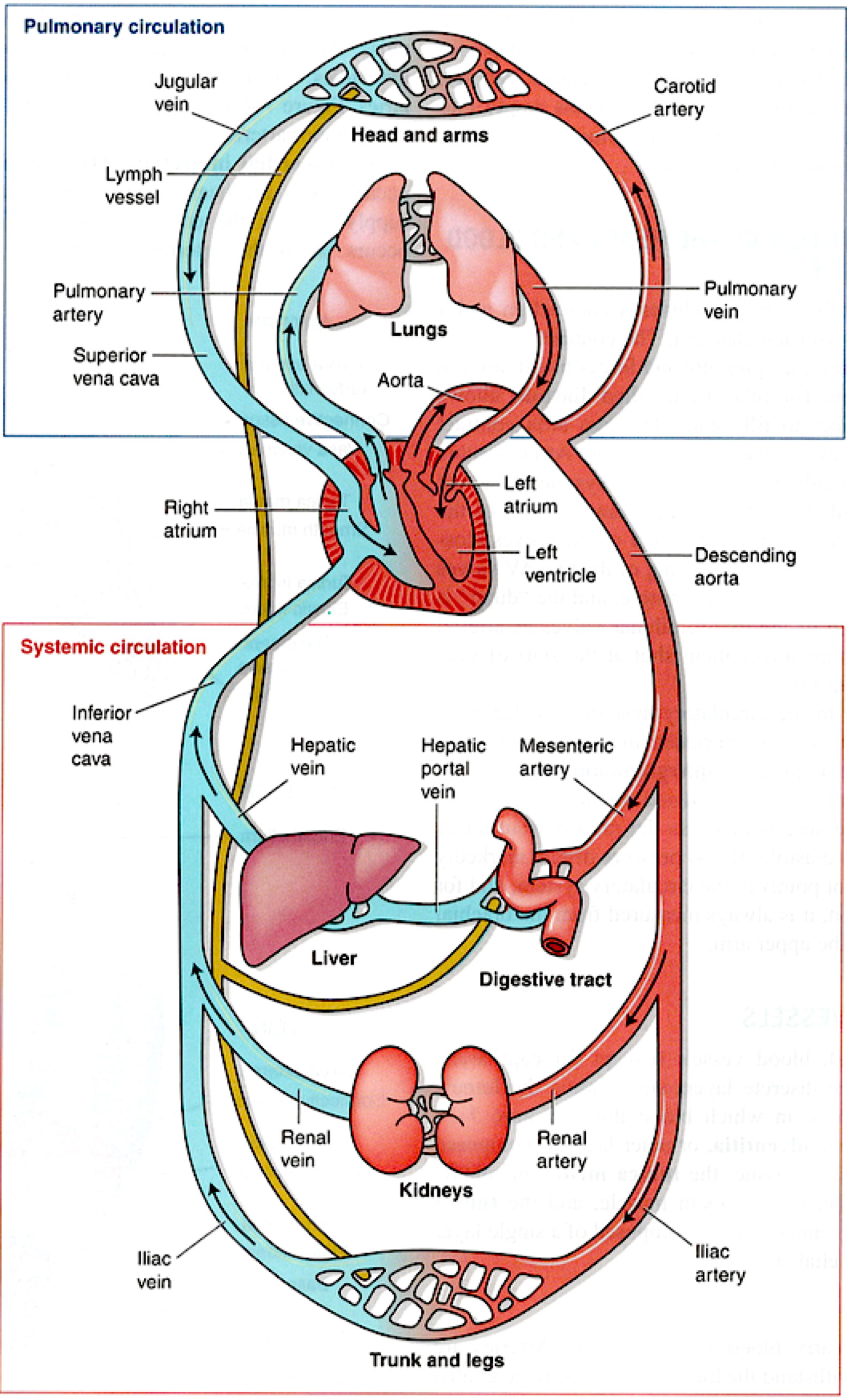
Pulmonary circulation
Carries blood from right ventricle of heart to lungs
Carbon dioxide is removed: oxygen is picked up
Oxygenated blood returns to the left atrium of the heart
Systemic circulation
Carries oxygenated blood and nutrients from left ventricle to body cells
Returns deoxygenated blood with carbon dioxide and wastes from cells to right atrium
A series of valves ensures that blood flows in the correct direction as it is pumped through the heart. The valves of the heart are made up of endocardium tissue.
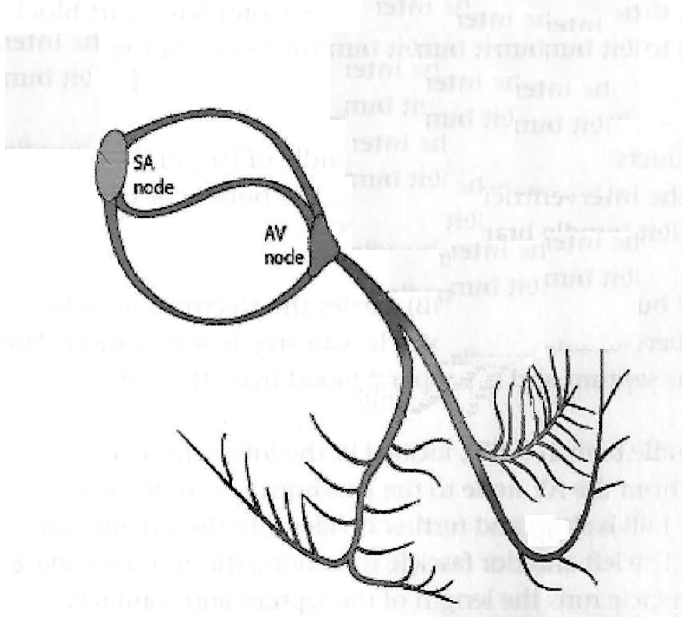
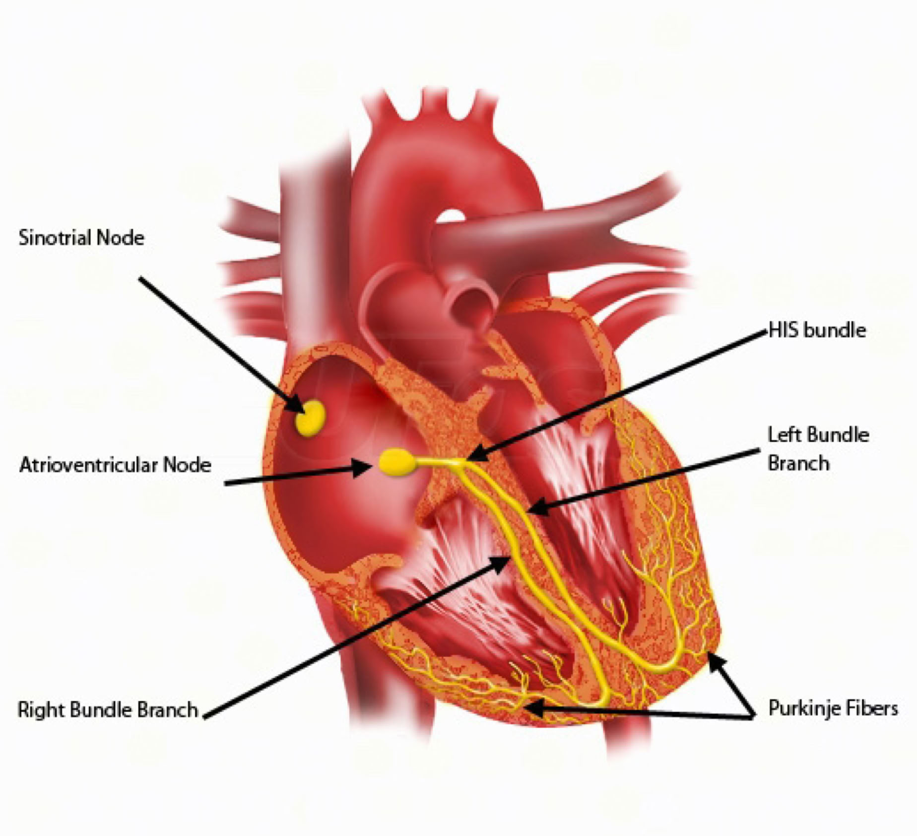
Conduction System
Normal pathways
The synchronous, rhythmic contraction of the heart muscle is controlled by an electrical conduction system.
In the normal heart, electrical impulses are initiated in the sinoatrial node (SA node), conducted through both atria, and directed to the atrioventricular node (AV node).
The AV node delays transmission of the signal to the ventricles, allowing them to completely fill with blood.
Depolarization continues toward the apex of the heart through the bundle of His and the left and right bundle branches until it terminates in the Purkinje fibers.
As the electrical impulses reach the myocardium, the muscle cells depolarize and contract.
In addition to the organized conduction system, the heart muscle can initiate its own impulse. This property is called automaticity.
Occasionally, a small area in atrial or ventricular tissue becomes irritated and initiates an electrical impulse from outside the normal pathways.
These are called ectopic beats and can be seen on the EKG as premature atrial, junctional, or ventricular complexes.
Sinoatrial node:
The SA node is found in the right atrium and functions as the primary pacemaker of the heart.
In adults, it fires approximately 60 to 100 times per minute. Depolarization of the right atrium occurs first, followed closely by the left.
The P wave seen on the EKG is a result of atrial depolarization.
Atrioventricular node: The atrioventricular (AV) node is the only part of the conduction system that connects the atria to the ventricles.
Just below the AV node lies the AV junction, where the AV node and the bundle of His come together.
The junction can function as a backup pacemaker if the SA node fails, and fires at approximately 40 to 60/min.
The AV node "holds" the electrical signal received from the SA node for a short period of time to allow the ventricles to completely fill with blood.
On the EKG, the time needed for an electrical impulse to travel from the SA node through the AV node to the ventricles is seen as the PR interval.
If the AV node is not conducting impulses normally, the PR interval may increase.
Complete heart block occurs when the AV node is unable to conduct any electrical impulses from the SA node to the ventricles.
Bundle of His:
The AV node conducts the impulse to the bundle of His, or interventricular bundle, located in the interventricular septum.
The bundle of His transmits impulses to the right and left bundle branches.
Right bundle branch:
The right bundle branch (RBB) carries the electrical impulse from the AV node to the Purkinje fibers of the right ventricle, causing depolarization.
Left bundle branch:
The left bundle branch (LBB), located in the interventricular septum, carries the electrical impulse from the AV node to the Purkinje fibers of the left
ventricle, causing depolarization.
Purkinje fibers:
The Purkinje fibers are a network of conduction pathways that
traverse the surface of the ventricles and depolarize them, initiating myocardial contraction.
In the absence of electrical stimulation from the SA and AV nodes, the Purkinje fibers will fire at an intrinsic rate of 20 to 40/min.