EKG Technician will perform several different types of EKG tests. The doctor will order a specific type of EKG based on the information needed to obtain about the patient’s cardiovascular system.
An electrocardiogram is a recording of the electrical activity of the heart.
The heart’s electrical activity prompts the muscle contraction that pump blood. The movement of electrical signals in the heart is called cardiac conduction.
Normal cardiac conduction starts in the upper part of the heart and moves in a downward pattern.
Conduction is continuous and occurs in cycles. Changes in the pattern can be normal or they can indicate a diseases or disorder of the heart.
The electrical activity recorded by the EKG is caused by changes in the heart’s cells.
2 Types of cardia electrical activity:
Depolarization: refers to change in electrical charge from negative to positive within the heart cells.
Depolarization causes the heart muscle to contract and pump blood.
Repolarization: refers to a change in electrical charge from positive back to negative.
Repolarization result in the heart muscle relaxing.
Each cardiac cycle is the beginning of a beat until the beginning of the next beat and consists of a depolarization phase and repolarization phase.
These electrical changes cause the atria and the ventricles to contract and relax at opposite times so that the chambers can fill and then pump blood.
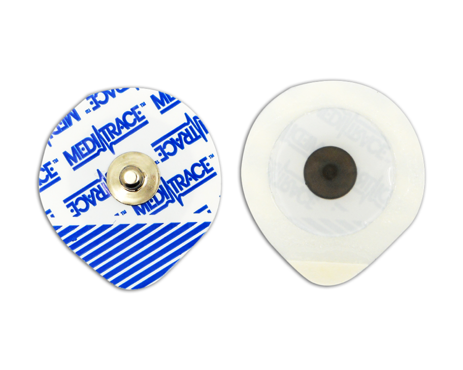
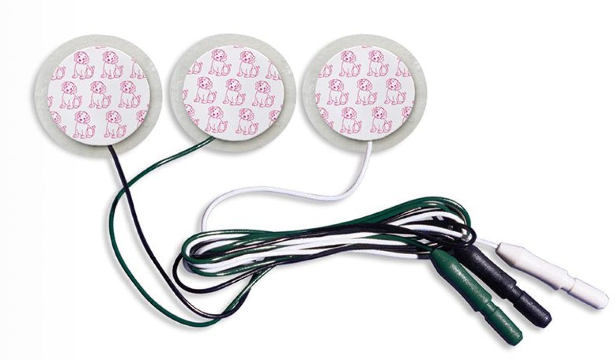
EKG machines are connected to patients using electrodes, or pads that conduct electricity.
Electrodes: are pads that transmit conductivity
Lead wires (or electrode cables): Transmit the heart’s electrical signals to the EKG machine.
The EKG machine measures the electrical activity of the heart in several directions called leads throughout a series of heartbeats.
Each lead provides a view of the heart based on the electrical activity between two or more electrodes.
The EKG records this activity on graph paper that is run though the machine at a fixed rate. The resulting record is called an EKG tracing.
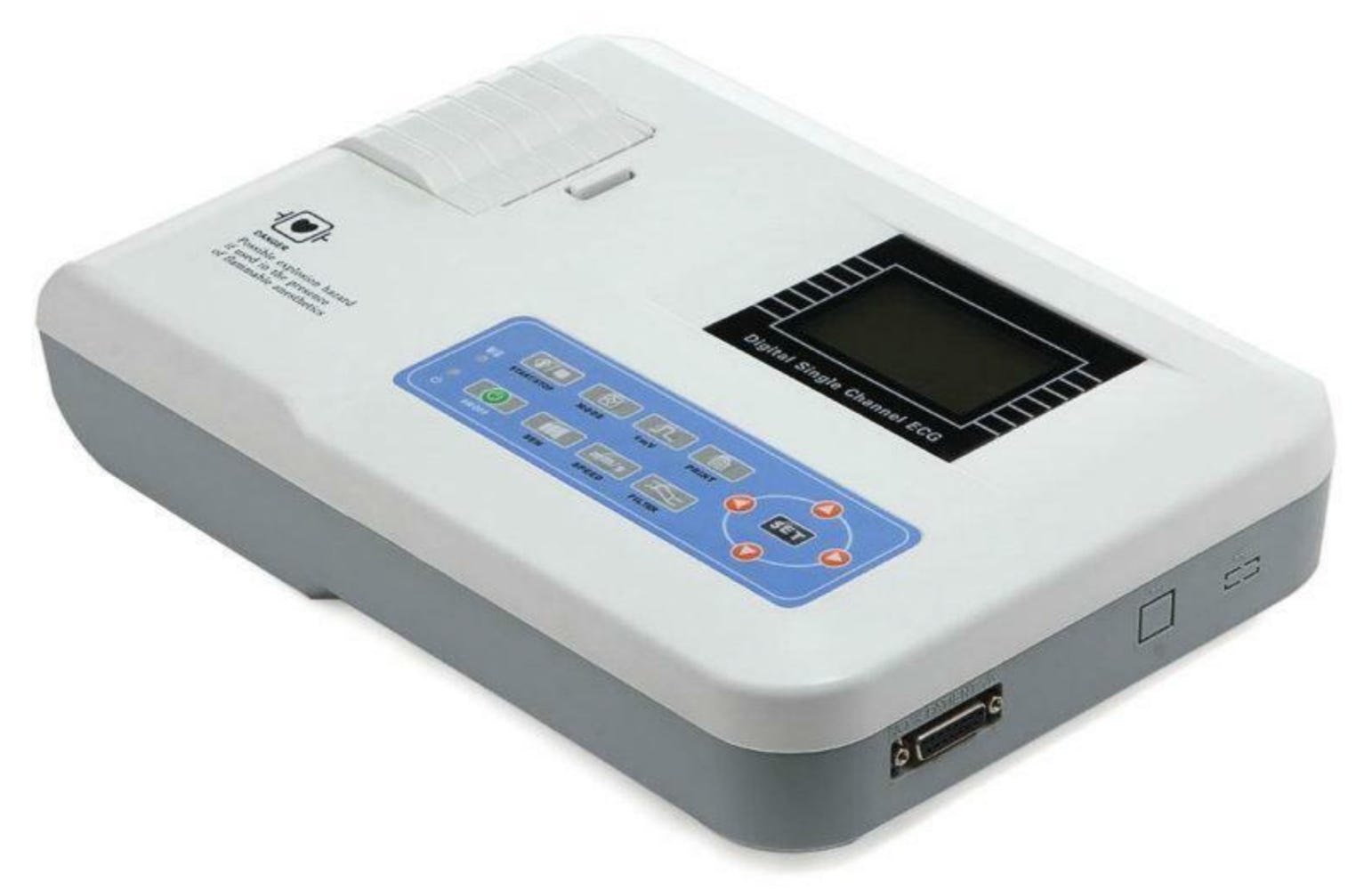
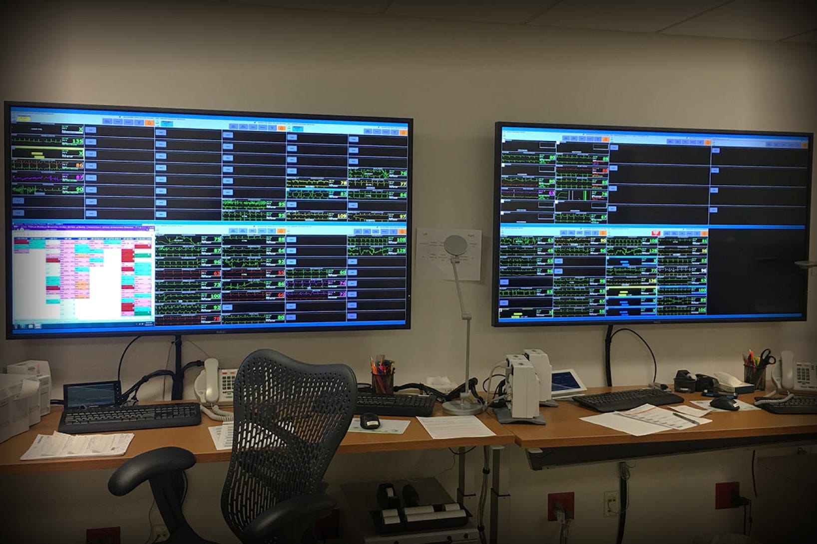
The Portable EKG Machine:
The portable EKG machine is a combined unit that includes a screen and printer. The unit has a long cable that branches into 10 lead wires.
Each wire attaches to an electrode that is placed in a specific place on the patient’s body to complete an electrical circuit with the EKG machine.
The machine automatically read and records the electrical activity from the different leads, which are created by various combinations of signals from two or more lead wires.
Electrodes attach to the lead wires using snaps or clips. They are placed on the patient’s chest and extremities in specific locations.
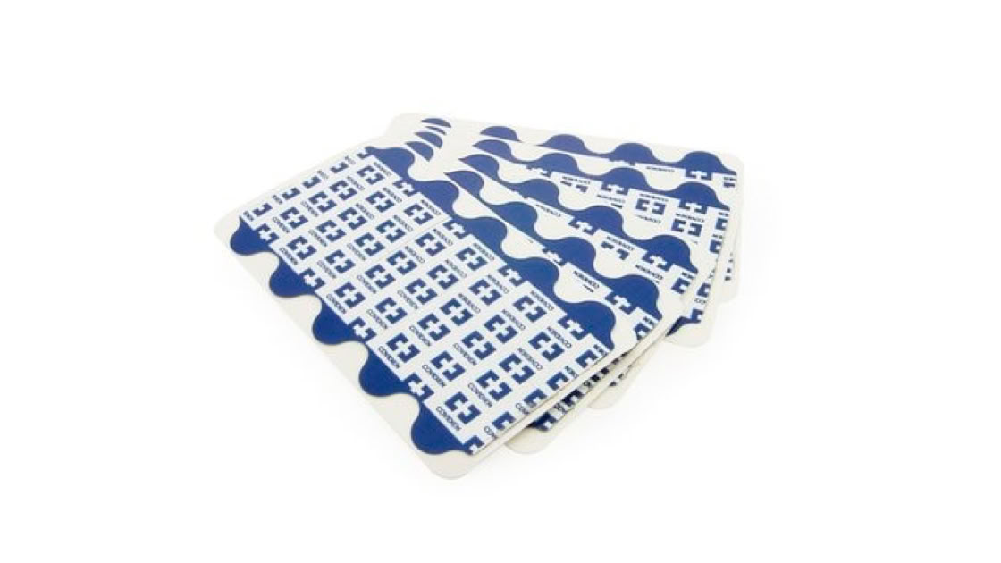
Different types of electrodes are used for different EKG tests. Thin, flat electrodes called resting electrode are usually used for routine diagnostic Screening EKG’s that require only one recording.
Circular pad electrodes called monitoring electrodes are used for continuous monitoring.
Electrodes contain a special gel to permit electrical conduction from the skin to the lead wire and on the EKG machine.
DIFFERENT TYPES OF EKG:
Continuous monitoring: Produces a long record of the heart’s activity. If the patient must be monitored on an EKG machine on an ongoing basis.
This type of monitoring is often done in emergency care settings when the nurse or doctor needs to be able to continuously assess the patient’s rate and rhythm.
Telemetry: Monitors heart rate at a remote monitoring station. Produced a longer record of the electrical activity to assist with the interpretation of any rhythm problem.
Monitoring is used in large facilities to monitor a patient’s EKG on an ongoing basis.
The patient wears a small device with five Lead wires. The device transmits information to a central monitoring station.
Specially trained healthcare workers view the patient’s cardiac activity at the central monitoring station and alert the nurse or doctor if the patient’s heart rate or rhythm changes.
Ambulatory monitoring: used when the doctor needs information about the heart over a period of time.
Holter monitors are one common type of ambulatory monitor.
These monitors are usually placed on the patient in a doctor’s office and removed after 24-48 hours.
The information stored in the machine is then downloaded and evaluated by the doctor.
Event monitors: only record the heart’s electrical activity when the patient presses a button to indicate that he is experiencing symptoms.
The most common EKG test for screening and diagnosis of the heart abnormalities is the 12-lead EKG. The 12 lead EKG has 10 cables.
The 12 lead electrocardiogram is called that because it takes 12 readings of the heart.
It is also used during stress tests, which monitors how the heart responds to physical activity.
The stress test evaluates the cardiac function under stress (induced by exercise or medication).
Identify the different EKG leads and lead groups
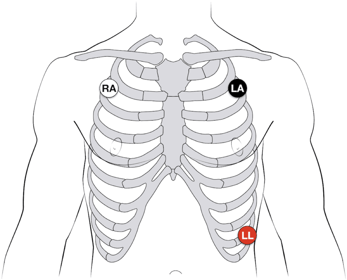
3-Lead EKG
The 3-lead EKG configuration is generally used to continuously monitor the patient's heart rhythm.
• White lead - Right shoulder or clavicle area
• Black lead -Left shoulder or clavicle area
• Red lead - Left lower abdominal area
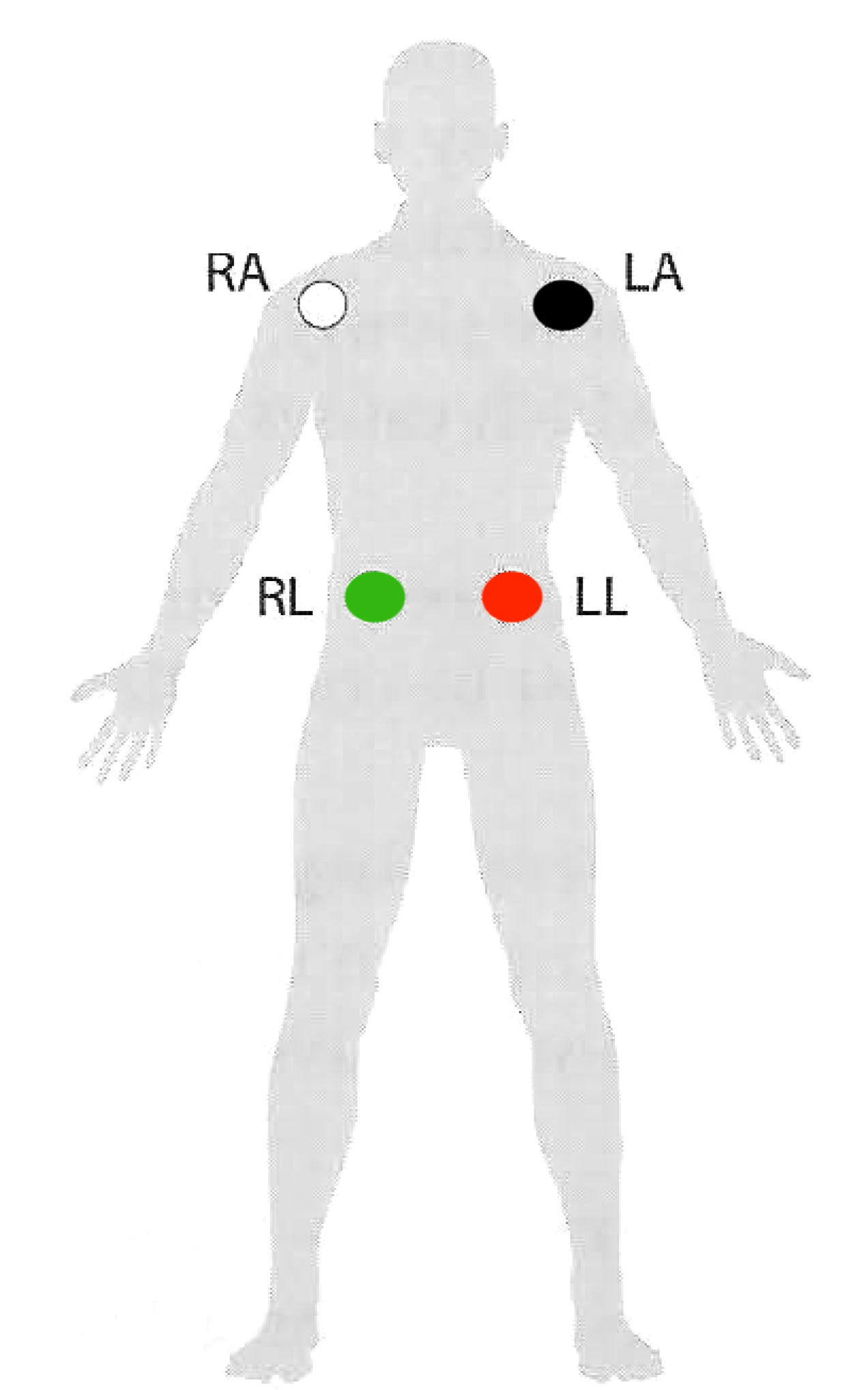
4-Lead EKG
The 4-lead EKG configuration is generally used to continuously monitor the patient's heart rhythm.
• White lead - Right shoulder or clavicle area
• Black lead -Left shoulder or clavicle area
• Red lead - Left lower abdominal area
• Green lead -Right lower abdominal area
5-Lead EKG
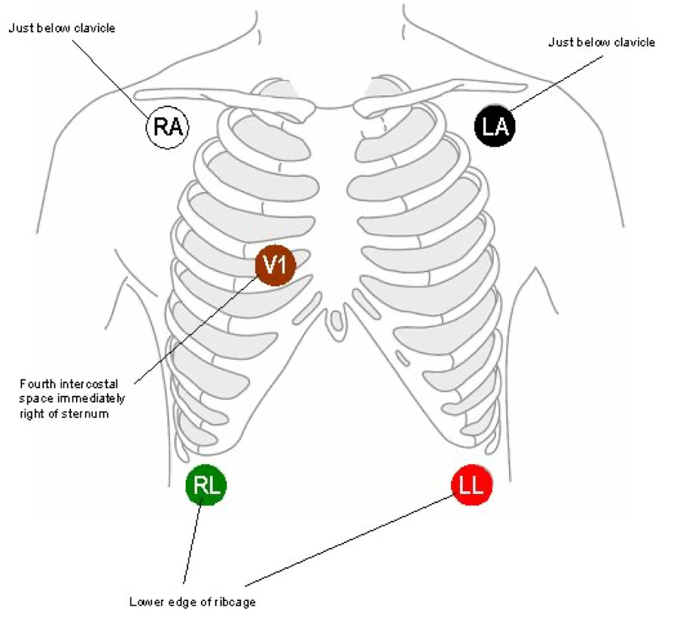
The 5-lead EKG configuration refers to the standard Holter monitor setup or the 5-lead rhythm monitor setup. The Holter monitor setup varies depending on the type of monitor. The 5-lead setup pictured here is also the most common configuration for Telemetry.
• White lead - Right sternum/clavicle area
• Black lead- Left sternum/clavicle area
• Red lead- Left lower thoracic area
• Green lead- Right lower thoracic area
• Brown lead- Just below and to the right
of the bottom of the sternum
12-lead
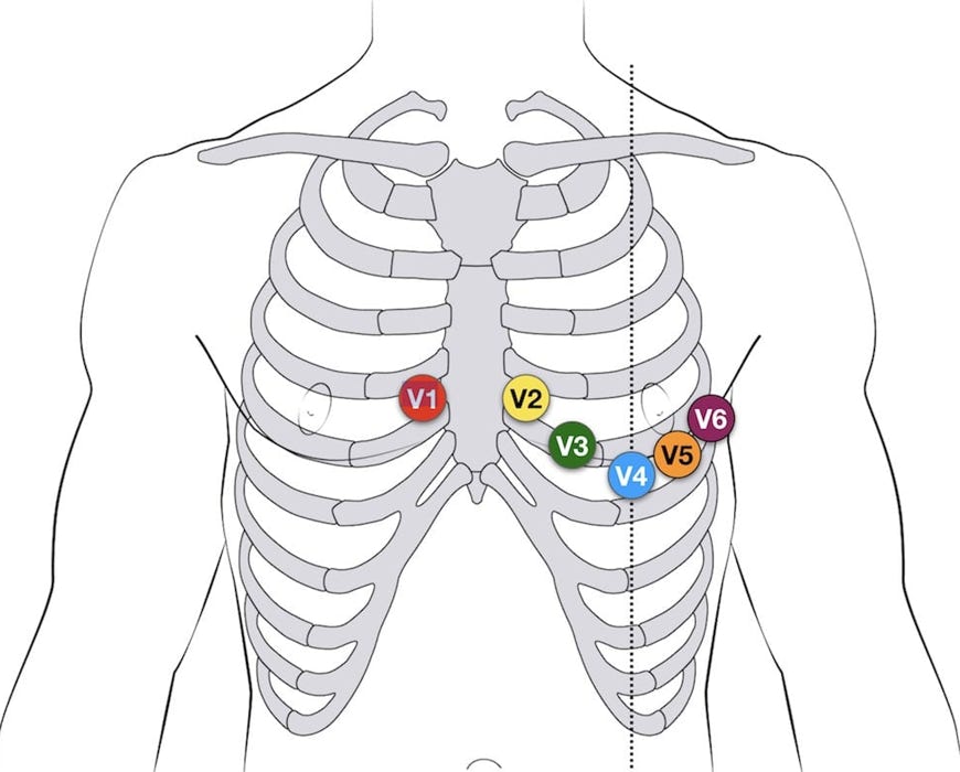
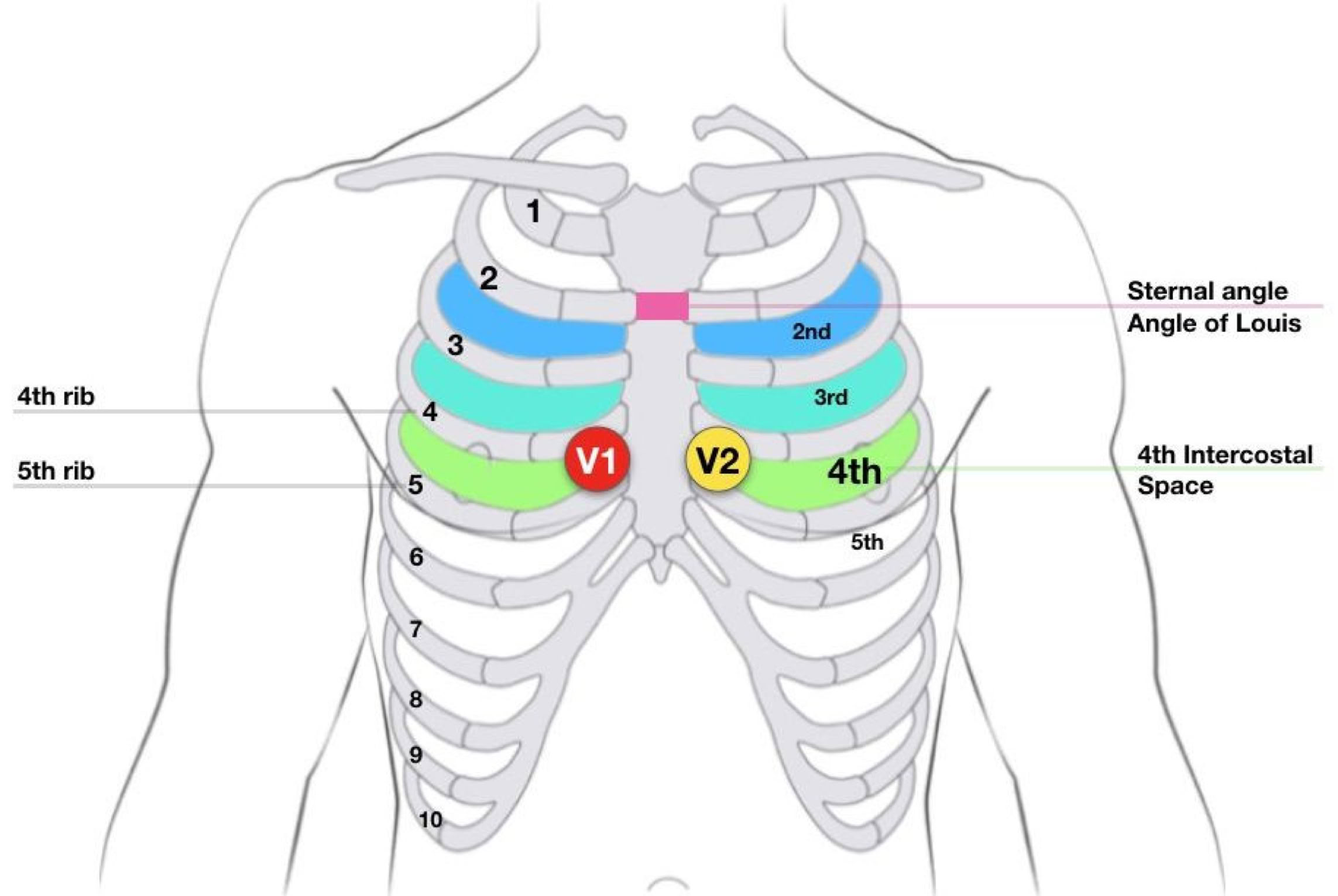
The standard 12-lead EKG requires placement of 10 electrodes; the standard 4 limb leads, plus 6 precordial leads.
Precordial Leads - Left side
• V1- 4th intercostal space R of sternum
• V2- 4th ICS, L of sternum
• V3 - between V2/V4
• V4- 5th ICS, midclavicular
• V5- 5th ICS between V4/V6
• V6- 5th ICS, midaxillary
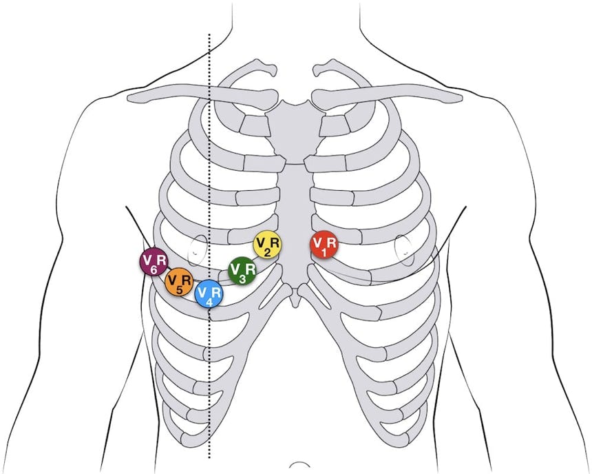
Certain conditions, including inferior wall ST segment elevation, Dextrocardia and pediatric patients less than 2 years old, require a right-sided 12-lead EKG. For patients who are ages 2-8, you can use either left- or right-sided EKGs. After age 8, you can no longer use the right-sided EKG.
You can also use the right-sided EKG for patients with certain conditions, including inferior wall ST segment elevation and myocardial infarction. Limb leads are placed in the normal fashion, but the precordial leads are placed as illustrated below.
Precordial Leads - Right side
• V1- 4th ICS, L of sternum
• V2- 4th ICS, R of sternum
• V3 - between V2/V4
• V4 - 5th ICS, midclavicular (R)
• V5- 5th ICS between V4/V6
• V6 - 5th ICS, midaxillary
Inferior wall infarction:
When there's an inferior wall infarction, you always need to consider posterior wall involvement. This diagram shows proper placement of leads V7, V8, and V9 for a posterior EKG. You will rarely perform the posterior EKG.
Amputation:
Lead placement must be modified in patients who have extremity amputation. Leads can be moved to an area just above the knees and elbows in patients who have distal extremity amputation. If the patient's amputation is close to the torso, EKG electrodes can be placed on the Posterior EKG- Electrode placement patient's torso.
LIMB LEADS OR BIPOLAR LEADS
All the lead used in a EKG test have names and are arranged into groups, which also have names.
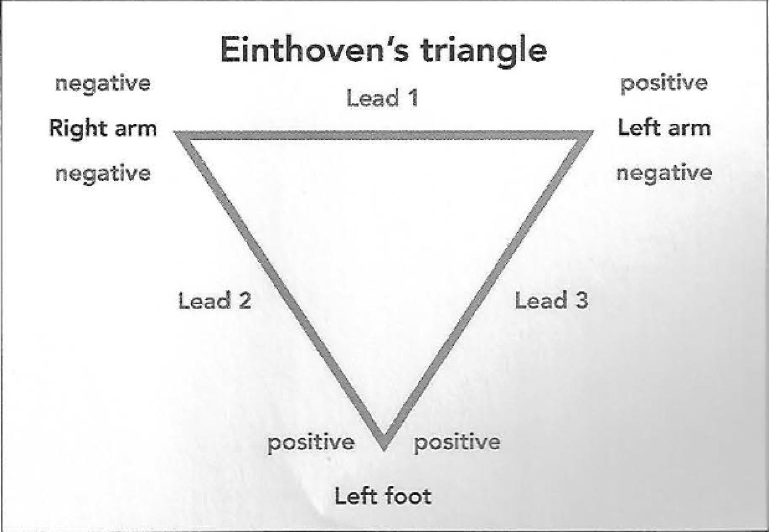
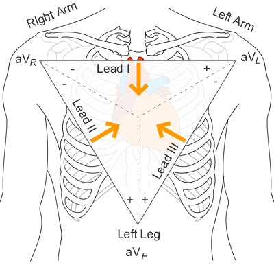
Leads I,II and III are called the limb leads or bipolar leads.
The limb lead electrodes form the point known as Einthoven’s triangle, named for the doctor who first recorded the electrical activity of the heart in the early 1900s.
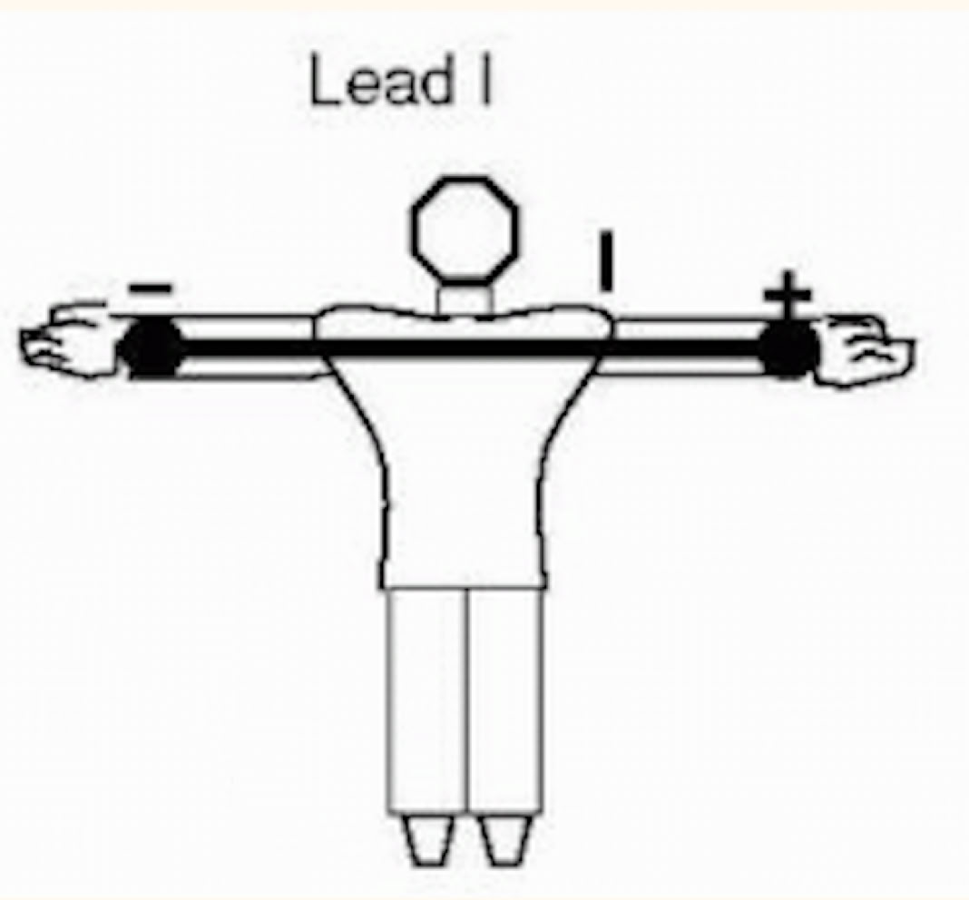
Lead 1 in the EKG measures the voltage between the left arm electrode (the positive pole) and the right arm electrode (the negative pole).
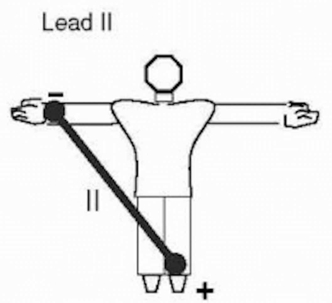
Lead II records the voltage between the left leg electrode (positive) and the right arm (negative).
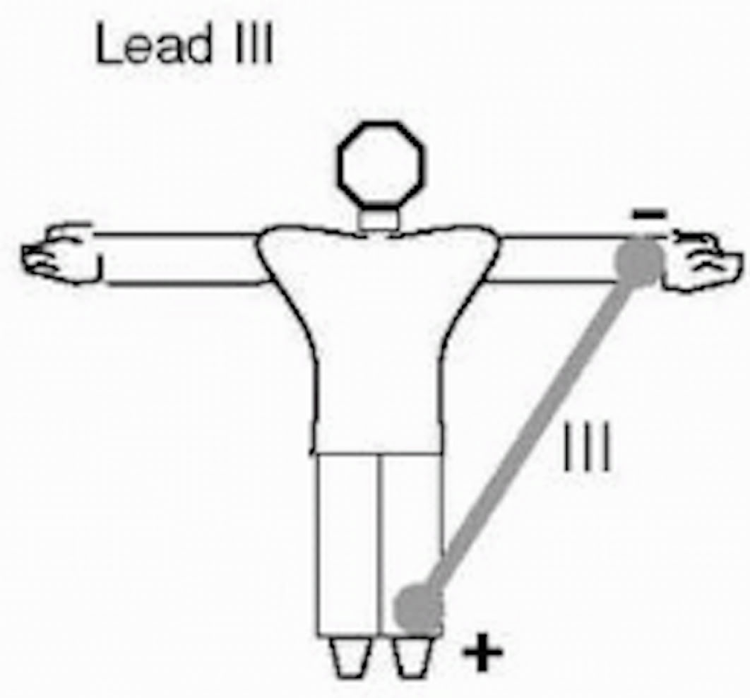
Lead III records the voltage between the left leg electrode (positive) and the left arm electrode (negative).
AUGMENTED OR UNIPOLAR LEADS
Leads aVR, aVL, and aVF are called augmented limb leads. This lead are unipolar leads. They focus on the electrical activity of one positive pole, measuring the flow of electrical current in one direction only.
Unlike the bipolar limb leads, which measures the flow of electrical current in both directions between a positive and negative pole, the augmented limb leads measure voltage at a positive pole in comparison to a neutral reference point. Neutral means without positive or negative electrical charge.
In the augmented limb leads the positive input is recorded at the right arm for aVR, the left arm for aVL, and the left leg for aVF. The right leg is considered the ground.
Wilson's central terminal:
Wilson's central terminal (WCT) is a reference point created by the three limb leads.
It serves as a reference point for six of the 12 leads, and serves as the "zero" end for each of the nine unipolar leads on the EKG.
Proper limb lead placement is critical to ensure proper calculation of WCT.
Improper limb lead placement creates an incorrect WCT reference point.
PRECORDIAL LEADS
Leads V1,V2,V3,V4,V5, and V6 are known together as the precordial leads, or chest leads. These leads are also unipolar.
The first step in placing the precordial lead is locate the angle of Louis (also known as the sternal angle).
To detect the angle of Louis, place your fingers at the base of the throat in the central position and move them down-ward, a bony lump will be felt, this is the angle of louis and represent the second intercostal space.